Category: Pathophysiology

pathophysiology

sickle cell anemia

Pathophysiology of Sickle cell anemia

chibueze uchegbu | December 12th, 2021


In the pathophysiology of Sickle cell anemia or Sickle cell disease (SCD), we learn that it is a systemic disorder caused by a mutation in the gene encoding the β chain of hemoglobin.

This mutation leads to the production of sickle hemoglobin HbS. Sickled red blood cells were first observed in 1910 by JB Herrick in a Black West Indian student.

Introduction to pathophysiology of sickle cell anemia

Sickle cell anemia or Sickle cell disease is the result of a single base-pair change, thymine for adenine, at the sixth codon of the beta gene.

in which the change encodes Valine instead of glutamine in the sixth position on the beta-globin molecule.

Beta globin haplotypes are the different DNA structures associated with the sickle gene are identified by a pattern of restriction enzyme sites.

Therefore, it is important to note that the HbS gene is prevalent in malaria-endemic regions, distribution is however worldwide, with the greatest incidence in tropical Africa.

sickle cell anemia

It occurs in the USA, Middle East, India, the Caribbean, South and Central America, Turkey, and throughout the Mediterranean region.

A 2009 study carried out in Nigeria showed that 2-3% of Nigerian newborns are homozygous.

Incidence in the general Nigerian population is 1:300 with Frequency of the S gene in Nigeria is about 25% Mode of inheritance= autosomal recessive.

Pathogenesis and pathophysiology of sickle cell anemia

The hemoglobin molecule (alpha and beta-globin subunits) picks up oxygen in the lungs and releases it when the red cells reach peripheral tissues, such as the muscles in Sickle cell anemia or Sickle cell disease.

And also, normal red cells maintain a basic disc shape, whether they are transporting oxygen or not but the case is different with sickled hemoglobin.

Sickle hemoglobin exists as isolated units in the red cells.

when they have oxygen bound, Whereas sickle hemoglobin releases oxygen in the peripheral tissues, however, the molecules tend to stick together and form long chains or polymers (polymerization).

These rigid polymers interact with the cell and cause it to bend out of shape Polymerized sickle hemoglobin does not form single strands.

But the molecules group in long bundles of 14 strands each that twist in a regular fashion, much like a braid.

In Sickle cell anemia or Sickle cell disease, most distorted cells are simply shaped irregularly, a few have a cresent-like appearance under the microscope.

These cresent-like or “sickle-shaped” red cells gave the disorder its name. A single red cell may traverse the circulation four times in one minute. Sickle hemoglobin undergoes repeated episodes of polymerization and depolymerization (sickle-unsickle cycle).

This cyclic alteration in the state of the molecules damages the hemoglobin and ultimately the red cell itself.
 Diameter of RBC =7microns
 Diameter of capillaries =3microns

Summary on the sickle cell anemia,


In
Sickle cell anemia or Sickle cell disease Polymers tend to grow from a single start site (nucleation site) and often grow in multiple directions. Star-shaped clusters of hemoglobin S polymers develop commonly.


Sickling occurs at the venous end of capillaries wh
ereas unsickling occurs at the arterial end.


Two essential pathological processes arise from sickling: Haemolysis and vaso-occlusion

The pathological features of SCD relate to the shortened life span of the sickled blood cells, (16-20 days rather than a lifespan of 120 days in normal red cells) which leads to hemolytic anemia

The mechanism is likely to be responsible for complications such as pulmonary hypertension and stroke.

Sickled cells directly cause Small vessel occlusion

sickle cell anemia

Vaso-occlusion is the major cause of morbidity and mortality in sickle cell anemia, accompanied by occlusion of blood vessels followed by ischemia or infarction in various tissues, leading ultimately to progressive end-organ damage.


The process of vaso-occlusion


It was first thought that hemoglobin S polymerization resulted in the entrapment of sickled, poorly deformable erythrocytes that mechanically blocked small-caliber vessels.

Damage similar to that seen in patients with the atherosclerotic vascular disease has been seen in the large cerebral vessels of patients with Sickle cell anemia or Sickle cell disease, including intimal hyperplasia, and fibroblast and smooth muscle proliferation.

It will be of great importance to consider these terms [CRISES]; Vaso-occlusive, Haemolytic, Aplastic, and Acute sequestration.


VASO-OCCLUSIVE CRISIS


This crisis is due to obstruction of blood flow in the smaller venules, capillaries and even in medium-sized or large arteries, as a result of the increased viscosity and sludging associated with sickling.

The increased adhesiveness of sickled reticulocytes worsens occlusion. Vaso-occlusion is the pathophysiologic basis of most of the clinical.


HAEMOLYTIC CRISIS


Hemolytic crisis makes RBCs are broken down at a more rapid rate than during the steady-state of the disease, it is precipitated by malaria and bacterial infections.

Features include:


*Severe anemia, Cardiac failure
*Jaundice, hepatomegaly.
*Encephalopathy-seizures, altered sensorium
*The terms ‘ hemolytic’ and ‘hyper haemolytic’s are often used interchangeably, but the latter technically refers to the co-existence of SCA and G6PD[glucose 6-phosphate dehydrogenase deficiency].
APLASTIC CRISIS

There is a shut down of the bone marrow which is usually limited to the red blood cell precursors, making the patient become profoundly anemic and may go into high-output cardiac failure.

Anemia reoccurs after blood transfusions until the crisis is over, Several viruses are associated with this syndrome most especially parvovirus B19.

ACUTE SEQUESTRATION


The Pooling of blood in the spleen is a frequent occurrence in children with sickle cell anemia, particularly in the first few years of life, resulting in splenic sequestration crisis.

They are often associated with viral or bacterial infections; acute chest syndrome occurred in 20% in one series.

The usual clinical manifestations are sudden weakness, pallor, tachycardia, tachypnea, and abdominal fullness.

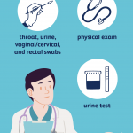
DIAGNOSIS OF SICKLE CELL ANEMIA


Clinical …….80% of cases.
Laboratory diagnosis:
Electrophoresis…cellulose acetate

DIAGNOSTIC METHODS INCLUDE:


Full blood count, blood film and reticulocyte count, bilirubin.
Hb electrophoresis using cellulose acetate or agar gel
High-performance
liquid chromatography (HPLC)
Isoelectric focusing (IEL)
Polymerase chain reaction (PCR)
Supplementary genetic tests

MANAGEMENT FOR A STEADY-STATE


Determine and record physical/Haematol Parameters.
Avoid factors that encourage sickling.
Folic acid supplementation.
Malaria prophylaxis.

Treatments

ANTISICKLING AGENTS

Hydroxycarbamide, formerly known as Hydroxyurea.

15mg/kg/24 hrs. gradually increase to max of 30mg/kg/24hrs. Monitor FBC, LFT and HbF. Increase in HbF is usually 10-15%
Trade name –Hydrea
*5-Azacytidine,
*decitibine
*Histone deacetylase inhibition: short-chain fatty acids –butyric acid.
*Recombinant human erythropoietin (rhEPO)


It is important to note that hydroxycarbamide is the most successful drug therapy for scd.

It is a cytotoxic and cytoreductase antimetabolite that acts via inhibition of DNA synthesis by inhibiting ribonucleotide reductase. Known pharmacological effects that may contribute to the drug’s efficacy in SCD include:

Hydroxycarbamide


1.increase in red cell content of Hb F.
2.dose related cytoreductase effects on neutrophils.
3.increase in water content of red cells.
4.increased deformability and successful microvascular navigation of sickled cells.
5.altered adhesion of RBCs to endothelium by decreasing the expression of endothelial adhesion molecules.
Treatment with hydroxycarbamide shows significant reduction in bone pain crises, admissions, and need for blood transfusion#

Gene therapy

The term gene therapy is applied to any maneuver in which genes or genetically modified cells are introduced into a patient for therapeutic benefit. Gene therapy corrects sickle cell disease in mice, scientists report therapies designed to treat genetic disease in humans.

The therapy transfers an anti-sickling variant of the faulty gene to the bone marrow, where it incorporates itself into the stem cells that give rise to red blood cells. In two mouse models, the new gene was rapidly expressed in 99 percent of all circulating red blood cells, preventing sickling and other signs of the disease, Leboulch said.

Recommendation

Pathophysiology of syphilis

Pathophysiology of fragile x syndrome in female

Pathophysiology of chlamydia

How to prevent skin to skin STDs; Best read

facts to know about astigmatism

Facts to Know About astigmatism

chibueze uchegbu | October 25th, 2021


Facts to Know About Astigmatism range from but are not limited to the Diagnosis Process and Treatment Options.

Astigmatism is one of the most prevalent eye conditions that affect millions of people around the world.

But what is astigmatism? Astigmatism is an eye condition wherein the cornea is irregularly shaped. The cornea is the transparent part of the eye that covers the front portion of the eye.

Together with the sclera (the white of your eye), it serves as a barrier against foreign bodies.

Introduction to facts to know about astigmatism

The normal cornea is round. However, for people with astigmatism, it is shaped irregularly and as a result, the eye has difficulties focusing light rays to one point and vision becomes out of focus at any distance.

This, in turn, causes blurry vision, discomfort, and headaches.

Additionally, the curvature of the cornea can change over time, resulting in an increase or decrease in astigmatism. This usually happens in adulthood and often precedes the growth of naturally occurring cataracts.

Early and correct diagnosis and treatment of astigmatism can correct and prevent this condition from worsening.

Additionally, there are many treatment options for this eye condition, including glasses and toric contact lenses for astigmatism.

Diagnosis of Astigmatism

If you have frequent headaches, experience blurred vision at any distance, fatigue, and eye strain, and need to squint to see clearly, you may already have astigmatism. You need to see a specialist immediately for a complete and proper diagnosis.

Once you have chosen your ophthalmologist, you will undergo a series of tests so that they can check your eye health.

These include:

●      Visual acuity

A test wherein you are asked to read letters on a Snellen eye chart or distance chart. You will then receive a measurement regarding how well you can see at a distance.

●      Refraction

Under this exam, the ophthalmologist will use different instruments and aim bright lights directly at your eyes as you look through several lenses. They will then determine how your eyes bend light.

●      Keratometry or topography

This procedure measures the curve of your cornea. Your specialist will focus light on the cornea and measure its reflection. You have to undergo this test if you want to use contact lenses to correct your astigmatism.

These tests will help your ophthalmologist understand the different aspects of your eyes and vision and determine the prescription you need so that you can see better with your eyeglasses or contact lenses.

Treatment Options

After your diagnosis, your ophthalmologist will discuss with you your treatment options and help you choose the best one.

Below are the usual options for correcting and improving astigmatism:

See below;

1.     Eyeglasses

Most people with astigmatism opt for eyeglasses to see more clearly.

These eyeglasses contain a special cylindrical lens prescription that makes the light bend into your eye properly. Some parts of the lenses have additional power to compensate for and correct astigmatism.

Depending on your diagnosis, your eyeglasses can also correct the refractive error you have, such as nearsightedness or farsightedness.

Your ophthalmologist may prescribe a single-vision lens that will give you clear vision at all distances. However, if you are 40 and have presbyopia, your specialist may recommend a bifocal or progressive addition lens.

2.     Contact lenses

Contact lenses provide clearer vision and a wider field of view, which is why many people prefer this treatment option over eyeglasses.

There are three types of contact lenses used for correcting astigmatism. These are:

●       Toric

Toric contact lenses are made with the same oxygen-permeable materials as standard lenses. However, they are designed in a way that they remain stabilized in the lens yet can still be rotated to the best position for consistently clear vision.

Toric lenses can also correct vision for people who are nearsighted and farsighted due to astigmatism.

Since they are contacts, they are more comfortable and convenient to wear than glasses.

●       Hybrid

Hybrid contacts combine the best features of toric and GP lenses. They have a wider diameter, with a firm central zone enclosed by soft material, such as hydrogel or silicone hydrogel soft lens material.

Due to its materials and design, hybrid contracts provide the comfort of soft toric lenses and the clear vision of GP lenses.

3.     Orthokeratology (Ortho-K)

Orthokeratology is a non-surgical procedure where a specialist inserts a series of gas-permeable contact lenses into the eyes to reshape the cornea.

The patient wears these contact lenses overnight. While asleep, the lenses gently and gradually reshape the corneas.

In the morning, the patient removes the lenses and will be able to see clearly throughout the day without wearing glasses or other types of contacts.

The effects of ortho-K are temporary, particularly during the early stages of treatment. Patients have to wear their lenses while sleeping every night for best results.

This treatment also does not improve vision permanently. If the patient stops wearing the lenses, their vision may return to its original condition.

4.     Refractive Surgery

Laser surgery is also used to change the shape of the cornea. After the procedure, you will have healthy eyes with no retina problems or corneal scars.

Laser-assisted in-situ keratomileusis or LASIK is the most popular laser eye surgery. Under this procedure, the specialist will make a thin, hinged flap in your cornea. They will then use an excimer laser to sculpt the shape of the cornea.

Once done, the surgeon repositions the flap.

LASIK causes less pain than other eye surgeries. Also, most patients get back the full function of their eyes and clearer vision within a few days.

Laser-assisted subepithelial keratectomy or LASEK is another type of refractive surgery that can help you correct your astigmatism.

But instead of making a flap in the cornea, the specialist will loosen the epithelium or the cornea’s thin protective cover with special alcohol. They will then use a laser to change the shape of the cornea. Afterward, they will reposition the loosened epithelium.

Because a thinner layer is affected during the procedure, the eyes are less vulnerable to damage or injury. This surgery is also usually recommended if the patient has a thin cornea and is not suitable for LASEK.

Conclusion on the facts to know about astigmatism

Astigmatism may be a prevalent eye disorder, but the various treatment options ensure you can have clearer vision regardless of your age and the severity of your condition.

I am confident you know the most important things about astigmatism and the clever approaches to treating and managing this eye condition.

we are open to suggestions, recommendations, and commendations, kindly air your view in the comment box below.

Recommendations

Best eye hospitals in Asia

Learn about Medical Tourism in Turkey

10 Best Eye Hospitals In New York

15 Best eye hospitals in India 

pathology,pathogens, what are the types of pathogens

What is a pathogen? best 4 main pathogens

Pharm. Somtochukwu | September 7th, 2021


What is a pathogen? The term pathogen came into use in the 1880s and was coined from the Greek word “pathos” and “genēs” which means “suffering” and “producer of” respectively, which means pathogen can be said to be a producer of suffering.

Typically, the pathogen is used to describe a disease-causing microscopic agent, such as a bacterium, virus, fungus, and parasite

What is a pathogen? The scientific study of microscopic organisms, including microscopic pathogenic organisms, is called microbiology, while the study of diseases that may include these pathogens is called pathology.

Introduction to What is a pathogen?

Pathogenic organisms have a single goal, which is to survive and multiply in their hosts.

Pathogens are specially adapted to infect a host, bypass the host’s immune responses, reproduce within the host, and escape its host for transmission to another host and therefore, disrupt the normal physiology of a multi-cellular animal or plant.

However, pathogens can infect unicellular organisms from all of the biological kingdoms.

There are several substrates and pathways whereby pathogens can invade a host.

What is a pathogen?

The human body contains many natural defenses against some of the common pathogens in the form of the human immune system and some “helpful” bacteria present in the human body’s normal flora.

Some pathogens are responsible for massive amounts of casualties and have had numerous effects on afflicted groups.

Today, many medical advances have been made to safeguard against infection by pathogens.

Social advances such as food safety, hygiene, and water treatment have reduced the threat from some pathogens.

What are the different types of Pathogens?

Pathogens vary in different ways and there are different pathogens with variable modes of infections. There are four main pathogens namely

  • Viruses
  • Bacteria
  • Fungi
  • Parasites

what is a pathogen?

Virus as a pathogen

A Virus is a non-living piece of genetic code, such as DNA or RNA, and protected by a coating of protein.

In the case of viral infection, viruses invade host cells within your body and then use the components of the host cell to replicate, producing more viruses.

After the replication cycle is complete, these new viruses are released from the host cell.

Some viruses can remain dormant for a time before multiplying again. When this happens, a person appears to have recovered from the viral infection but gets sick again.

Antiviral drugs are used for viruses while Antibiotics do not kill viruses and therefore are ineffective as a treatment for viral infections.

Bacterium as a pathogen

A Bacterium is a unicellular microorganism. It is diverse and has a variety of shapes and features, and has the ability to live in just about any environment, including in and on your body. It is essential to note that not all bacteria cause infections. The bacteria with the potential to induce infections are said to be pathogenic.

Antibiotics are used to treat bacterial infections. Some strains of bacteria have become resistant to antibiotics, making them difficult to treat. This can happen naturally but also happens because of the overuse of antibiotics, according to the World Health Organization.

Fungus as a pathogen

A Fungus cell has a nucleus and other components protected by a membrane and a thick cell wall. Their structure can make them harder to be eradicated.

Some new strains of fungal infections are proving to be especially dangerous, such as Candida aureus, and have prompted more research into fungal infections.

what is a pathogen?

Parasite as a pathogen

Parasites are organisms that behave like tiny animals, living in or on a host and feeding from or at the expense of the host.

Though parasitic infections are more common in tropical and subtropical regions, they can occur anywhere.

Three main types of parasites can cause disease in humans. These include:

  • protozoa, which are single-celled organisms that can live and multiply in your body
  • helminths, which are larger, multi-celled organisms that can live inside or outside your body and are commonly known as worms
  • ectoparasites, which are multi-celled organisms that live on or feed off your skin, including some insects, such as ticks and mosquitos “biologically known as vector while the plasmodium transmitted by mosquitoes is a protozoan”

They can spread in several ways, including through contaminated soil, water, food, and blood, as well as through sexual contact and via insect bites.

Different diseases caused by different pathogens

Viruses, bacteria, fungi, and parasites cause a variety of infections, which we will be looking at in this article

Virus as a pathogen and some of the infections it causes

Viruses can cause a number of infections, many of which are contagious. Examples of viral diseases include:

Bacterium as a pathogen and some of the diseases it causes

  • tuberculosis
  • cellulitis
  • cholera
  • gonorrhea

Fungus as a pathogen and some of the diseases it causes

  • ringworm
  • thrush
  • athlete’s foot

Parasite as a pathogen and some of the diseases it causes

  • malaria
  • giardiasis
  • intestinal worms

Routes of transmission of pathogens

Pathogens are transmitted either directly or indirectly.

Direct transmission involves the spread of pathogens by direct body-to-body contact.

This type of direct transmission (mother-to-child) is also known as vertical transmission.

Pathogens can also be spread by indirect transmission, which involves contact with a surface or substance that is contaminated with pathogens. It also includes contact and transmission through an animal or an insect vector.

Types of indirect transmission include:

  • Airborne 

The pathogen is expelled (typically by sneezing, coughing, laughing, etc.), remains suspended in air, and is inhaled by or comes in contact with the respiratory membranes of another person

  • Droplets 

Pathogens are contained in droplets of body fluid (saliva, blood, etc.) contact another person or contaminate a surface. Saliva droplets are most commonly spread through sneezing or coughing

  • Foodborne 

Transmission occurs through eating contaminated food or improper cleaning habits after handling contaminated food.

  • Waterborne 
The pathogen is spread by consumption or contact with contaminated water.
  • Zootonic 

The pathogen is spread from animals to humans. This includes insect vectors that transmit disease through biting or feeding and transmission from wild animals or pets to humans.

what is a pathogen?

While there is no way to completely prevent pathogen transmission, the best way to minimize the chances of acquiring a pathogenic disease is by maintaining good hygiene.

These include

  • Wash your hands often both during indoors or outdoors activities
  • Get vaccinated and ensure vaccinations are up to date
  • Prepare, cook, and store meat and other food items properly
  • Avoid sharing drinking glasses or utensils
  • Protect against insect bites
  • Travel wisely by getting informed about health risks and special vaccinations
  • Practice safe sex.
pathophysiology of scabies

Pathophysiology of scabies

chibueze uchegbu | July 18th, 2021


The pathophysiology of scabies is worthwhile and everyone ought to know the cardinal symptoms and how to prevent them.

Oftentimes people of all races and backgrounds are affected by the infestation of sarcoptes scabiei mite, as well as the human itch mite worldwide, this infestation is generally known as scabies.

Sarcoptes scabiei,  falls under the class of Arachnida and infect both human and animal at varying degrees.

Introduction to the pathophysiology of scabies

A readily treatable infestation, scabies remains common primarily because of diagnostic difficulty, inadequate treatment of patients and their contacts, and improper environmental control measures. Scabies is a great clinical imitator.

Its spectrum of cutaneous manifestations and associated symptoms often results in delayed diagnosis. In fact, the term “7-year itch” was first used with reference to persistent, undiagnosed infestations with scabies

In humans, the availability of mites will determine how contagious the infestation will be.

for instance, incrusted or Norwegian scabies {because it was first analyzed in Norway} is highly contagious due to thousands of these microscopic mites present. The availability of these mites is because of a compromised immune system like in the case of HIV/AIDS.

Pathophysiology of scabies

The pathogen responsible for scabies is sarcoptes scabiei mite. This infectious agent burrows under the skin using its mouth and the sophisticated front legs, the male and female act synergistically.

When a fertile female finds its way under the skin, as it burrows it lays eggs which later hatch to larva. The larva attacks the hair follicle under the skin and equally makes the skin vulnerable to opportunistic dermatological diseases.

The movement of the mites brings about the itching sensation which gets worsens in the presence of eggs. The pathology is more mechanical than chemical.

Cardinal symptoms of scabies

Unrelenting Itching is brought about by the movement of the mite under the skin.

Uneasy scratching of the affected part might equally make the skin vulnerable to secondary infections like dermatitis. Reddish rashes of varying sizes are equally pronounced.

Others may include;

The hotness of the affected area `and Sores

The part of the body, mostly affected by this parasitic mite are the hidden areas of the body and joints like the in between the fingers, armpit, elbow, wrist, knee, genitalia, and gluteus.

The contraction of scabies is by direct contact with an infected person or by using the same bedding with an infected fellow.

Types of scabies

  • Typical scabies
  • Nodular
  • Crusted

Typical scabies on the pathophysiology of scabies

Typical or classic scabies is the most common. It causes an itchy rash on the hands, wrists, and other common spots. However, it doesn’t infest the scalp or face.

Classic scabies has a distribution involving the axillae, elbow flexures, wrists and hands, and genital area. In infants and small children, burrows are commonly located on the palms and soles.

One- to 3-mm erythematous papules and vesicles are seen in typical distributions in adults. The vesicles are discrete lesions filled with clear fluid, although the fluid may appear cloudy if the vesicle is more than a few days old.

Nodular scabies on the pathophysiology of scabies

as the name implies this type of scabies may develop as itchy, raised bumps, or lumps, it may also present as persistent nodules for months even after specific treatment of scabies. It oftentimes appears around the genital areas, groin or armpit.

Crusted scabies on the pathophysiology of scabies

pathophysiology of scabies

Some people with scabies may develop another form of scabies known as Norwegian scabies or crusted scabies. This is a more severe and extremely contagious type of scabies. People with crusted scabies develop thick crusts of skin that contain thousands of mites and eggs which usually arise from individuals that are immune-compromised.

How can one prevent the spread of Scabies?

The best way to prevent getting scabies is to avoid direct skin-to-skin contact with a person known to have scabies.

It’s also best to avoid unwashed clothing or bedding that’s been used by a person infected with scabies. Make sure you wash all your bedding materials in hot water that reaches 122°F (50°C).

These items should then be dried in the dryer on very high heat for at least 10 to 30 minutes.

It will interest you to read

4 Facts on the Pathophysiology of chlamydia

Pathophysiology of gonorrhea over 5 most important things to know

Pathophysiology of Sickle cell anemia 

Meningitis pathophysiology, best read of 2023

types of sexually transmitted diseases orinfections

10 Types of sexually transmitted diseases

Pharm. Somtochukwu | July 4th, 2021


The types of Sexually transmitted diseases (STDs) are ill-health conditions passed from one person to another through sexual activities.

You can contract an STD by having unprotected vaginal, anal, or oral sex with an infected person.

An STD has also been labeled a sexually transmitted infection (STI) or venereal disease (VD). Depending on the specific STD, infections may also be transmitted through other routes asides from sexual activities like breastfeeding and an unhygienic environment. 

HIV has other routes of transmission. For example, this STI can spread through the use of unsterilized drug needles.

Some STIs are benign, but others can lead to severe complications if the infected person does not seek treatment.

Introduction to the types of sexually transmitted diseases

Anyone can contract an STI, regardless of his or her sexual orientation and hygiene standards. Many STIs can transmit through non-penetrative sexual activity.

If you have one STI, it can often increase your chances of contracting another. Some STIs can also lead to severe consequences if left untreated. In rare cases, untreated STDs may even be fatal.

Fortunately, most STDs can be treated and be cured totally. Is essential to note that early and effective treatment can help relieve symptoms, lower your risk of complications, and protect sexual partners.

Symptoms of types of sexually transmitted diseases

  • Headaches
  • Difficulty swallowing
  • Discharge
  • Fever
  • Night sweats
  • Fatigue/weakness
  • Appetite loss
  • Weight loss
  • Chronic diarrhea
  • Nausea/vomiting
  • Itchy, rashy skin/skin lesions
  • Chronic coughing
  • Restlessness
  • Confusion/delirium
  • Difficulty breathing

Is essential to note that different sexually transmitted infections present different symptoms and there may be variations in gender

Some specific symptoms of STIs in men

Is important you note that men may contract sexually transmitted infection without a possible symptom although some common symptoms are:

  • Painful or swollen testicles
  • Pain or discomfort during sex or urination
  • Sores, bumps, or rashes on or around the penis, testicles, anus, buttocks, thighs, or mouth
  • Unusual discharge or bleeding from the penis

Some specific symptoms of STIs in women

In many cases, STDs don’t cause noticeable symptoms. When they do, common STD symptoms in women include:

  • Pain or discomfort during sex or urination
  • Sores, bumps, or rashes on or around the vagina, anus, buttocks, thighs, or mouth
  • The unusual odor from the vagina
  • Unusual discharge or bleeding from the vagina
  • Itchiness in or around the vagina

In this article, some common STIs are discussed to help you cruise in the pool of ideal health

Types of sexually transmitted diseases

SYPHILIS

Syphilis is among the types of sexually transmitted diseases however, it is a bacterial infection caused by the bacterium Treponema pallidum which slips into the bloodstream and is eventually sent to organs outside the reproductive tract. It often goes unnoticed in its early stages.

The first symptom to appear is a small round sore, known as a chancre. Since syphilis chancres aren’t painful and typically heal within four to six weeks, most boys don’t see a doctor. They assume the mysterious sore is gone for good, but one-third of men and women exposed to primary syphilis progress to a secondary infection

This sexually transmitted disease can develop on your genitals, anus, or mouth.  It’s painless but very infectious.

Later symptoms of syphilis can include:

  • rash
  • fatigue
  • fever
  • headaches
  • joint pain
  • weight loss
  • hair loss

If left untreated, late-stage syphilis can lead to:

  • loss of vision
  • loss of hearing
  • loss of memory
  • mental illness
  • infections of the brain or spinal cord
  • heart disease
  • death

Fortunately, if caught early enough, syphilis is easily treated with antibiotics. However, syphilis infection in a newborn can be fatal. That’s why it’s important for all pregnant women to be screened for syphilis.

The earlier syphilis is diagnosed and treated, the less damage it does. 

Types of sexually transmitted diseases

Human Immunodeficiency Virus (HIV)

HIV can damage the immune system and raise the risk of contracting other viruses or bacteria and certain cancers. If left untreated, it can lead to stage 3 HIV, known as AIDS. But with today’s treatment, many people living with HIV don’t ever develop AIDS when proper medications are taken.

AIDS is caused by the human immunodeficiency virus (HIV), which is spread through unprotected sexual intercourse with an infected person or through using a contaminated needle to inject drugs. It is also among the known different types of sexually transmitted diseases.

It can also be spread through intravenous drug use and much less commonly, blood, blood products, needles, or other sharp instruments contaminated with infected body fluids or blood.

In the early or acute stages, it’s easy to mistake the symptoms of HIV with those of the flu. For example, the early symptoms can include:

  • fever
  • chills
  • aches and pains
  • swollen lymph nodes
  • sore throat
  • headache
  • nausea
  • rashes

These initial symptoms are typically clear within a month or so. From that point onward, a person can carry HIV without developing serious or persistent symptoms for many years. Other people may develop nonspecific symptoms, such as:

  • recurrent fatigue
  • fevers
  • headaches
  • stomach issues

HIV is one of the deadliest sexually transmitted diseases because there’s no cure for it yet. Although, treatment options are available to manage it. Early and effective treatment can help people with HIV live as long as those without HIV.

Proper treatment can also lower your chances of transmitting HIV to a sexual partner. In fact, treatment can potentially lower the amount of HIV in your body to undetectable levels.

With recent advancements in testing and treatment, it’s possible to live a long and healthy life with HIV. 

Gonorrhea

Gonorrhea usually begins in the urethra (bladder opening) or the cervix. However, the rapidly proliferating Neisseria gonorrhoea bacterium is the pathogen that causes gonorrhea. It can migrate to the uterus and the fallopian tubes, giving rise to pelvic inflammatory disease (PID). The infection, like chlamydia, may also involve the rectum. It is also known as “the clap.”

Many people with gonorrhea develop no symptoms. But when present, symptoms may include:

  • a white, yellow, beige, or green-colored discharge from the penis or vagina
  • pain or discomfort during sex or urination
  • more frequent urination than usual
  • itching around the genitals
  • sore throat

If left untreated, gonorrhea can lead to:

  • infections of the urethra, prostate gland, or testicles
  • pelvic inflammatory disease
  • infertility

Symptoms typically occur two to ten days after exposure.

Men:

  • Penile discharge
  • Mild to a severe burning sensation when urinating
  • Can progress to epididymitis

Women:

  • Painful or burning sensation when urinating and/or yellow or bloody vaginal discharge
  • Abdominal pain
  • Bleeding between menstrual periods
  • Vomiting
  • Fever
  • Progression to pelvic inflammatory disease

Rectal Infection:

  • Anal discharge
  • Anal itching
  • Painful bowel movements

It’s possible for a mother to pass gonorrhea onto a newborn during childbirth. When that happens, gonorrhea can cause serious health problems in the baby. That’s why many doctors encourage pregnant women to get tested and treated for potential STDs.

Types of sexually transmitted diseases

Herpes

Among the types of sexually transmitted diseases is Genital herpes. It is also commonly called “herpes,” is a viral infection by the herpes simplex virus (HSV). It is transmitted through intimate contact with the mucous-covered linings of the mouth or the vagina or the genital skin.

The virus enters the linings or skin through microscopic tears. Once inside, the virus travels to the nerve roots near the spinal cord and settles there permanently.

When an infected person has a herpes outbreak, the virus travels down the nerve fibers to the site of the original infection. When it reaches the skin, the typical redness and blisters occur. After the initial outbreak, subsequent outbreaks tend to be sporadic. They may occur weekly or even years apart.

Two types of herpes viruses are associated with genital lesions: herpes simplex virus-1 (HSV-1) and herpes simplex virus-2 (HSV-2).

HSV-1 more often causes blisters of the mouth area while HSV-2 more often causes genital sores or lesions in the area around the anus.

The outbreak of herpes is closely related to the functioning of the immune system. Women who have suppressed immune systems, because of stress, infection, or medications, have more frequent and longer-lasting outbreaks.

Genital herpes is spread only by direct person-to-person contact. It is believed that a majority of sexually active adults carry the herpes virus.

Symptoms of genital herpes

Once exposed to the virus, there is an incubation period that generally lasts 3 to 7 days before a lesion develops. During this time, there are no symptoms and the virus cannot be transmitted to others. An outbreak usually begins within two weeks of initial infection and manifests as an itching or tingling sensation followed by redness of the skin.

Finally, a blister forms. The blisters and subsequent ulcers that form when the blisters break are usually very painful to touch and may last from 7 days to 2 weeks.

HPV (human papillomavirus)

Human papillomavirus (HPV) is a virus that can be passed from one person to another through intimate skin-to-skin or sexual contact. There are many different strains of the virus. Some are more dangerous than others. Nearly every sexually active person will have HPV at some point.

Most types of HPV have no symptoms and cause no harm, and your body gets rid of them on its own. But some of them cause genital warts.

Others infect the mouth and throat. Still, others can cause cancer of the cervix, penis, mouth, or throat.

The CDC recommends young women and men ages 11 to 26 get vaccinated for HPV. Since there’s no treatment for HPV.

A Pap smear can show most cervical cancers caused by HPV early on. HPV is also among the types of sexually transmitted diseases.

Chlamydia

Chlamydia results from an infection with Chlamydia trachomatis. It is a common infection that can spread through anal, vaginal, and oral sex. A pregnant woman can also transmit it to the baby during delivery.

It can infect the urethra (bladder opening) and cervix (uterus opening). It is common in youngsters aged fifteen to nineteen.  It is known to be the most prevalent bacterial STI in the United States.

chlamydia does not usually produce symptoms, but it can result in infertility and other complications if a person does not receive treatment for it. The disease is easily treated, but like other sexually transmitted infections, chlamydia tends to be silent and therefore go undiagnosed until it becomes more serious than in its early stages

If symptoms do occur, they may include a change in vaginal discharge and burning pain during urination.

Chlamydia which is among the types of sexually transmitted diseases, can also affect the rectum, if the infection occurs as a result of anal sex or if the infection spreads from another area. This can lead to:

  • rectal pain
  • rectal bleeding
  • rectal discharge

In those who do develop symptoms, these will usually appear 7–21 days after exposure to the bacterium.

types of sexually transmitted diseases and infections

Scabies among the types of sexually transmitted diseases

Oftentimes people of all races and backgrounds are affected by the infestation of sarcoptes scabiei mite, as well as the human itch mite worldwide, this infestation is generally known as scabies.

Sarcoptes scabiei,  falls under the class of Arachnida and infect both human and animal at varying degrees.

In humans, the availability of mites will determine how contagious the infestation will be.

for instance, incrusted or Norwegian scabies {because it was first analyzed in Norway} is highly contagious due to thousands of these microscopic mites present. The availability of these mites is because of a compromised immune system like in the case of HIV/AIDS.

Crabs, or pubic lice

Pediculosis pubis is an infection of the genital area caused by the crab louse (Phthirus pubis).

The lice (commonly called crabs) are small bugs that are visible to the naked eye without the aid of a magnifying glass or microscope. Crabs, or pubic lice, usually attach to pubic hair. Sometimes, however, they can affect the hair in the armpits, mustache, beard, eyelashes, or eyebrows.

The treatment for pubic lice is usually with a 1% cream rinse of permethrin that is applied to the affected area and washed off after 10 minutes.

The first stage in the life cycle will be the appearance of the eggs, which lasts 6–10 days. After hatching, the lice will look like tiny crabs. They need blood to survive and will live for around 2–3 weeks. In the last day or two, the females will lay more eggs, and the cycle will continue.

Pubic lice can spread from person to person during close physical contact, including sexual contact. They can also transmit via shared towels or bed linen like in the case of scabies.

Hepatitis B

Hepatitis B is a potentially life-threatening liver infection caused by the hepatitis B virus (HBV). It is among the types of sexually transmitted diseases rated as a major global health problem. It can cause chronic infection and puts people at high risk of death from cirrhosis and liver cancer.

A safe and effective vaccine that offers 98-100% protection against hepatitis B is available. Preventing hepatitis B infection averts the development of complications including the development of chronic disease and liver cancer.

Once a person has the virus, it can remain in their semen, blood, and other bodily fluids.

Transmission is possible through:

  • sexual contact
  • using nonsterile equipment for injections
  • puncturing the skin with a sharp object where the virus is present

it can also be congenital although, as long as the nipples are not cracked, the risk of transmitting the virus through breast milk is negligible, according to the Centers for Disease Control and Prevention (CDC).

Trichomoniasis

Trichomoniasis, or trich, is caused by Trichomonas vaginalis it can affect both males and females, but females are more likely to experience symptoms

More women than men get trichomoniasis, which is caused by this tiny parasite.

Men and women can give it to each other through penis-vagina contact. Women can give it to each other when their genital areas touch.

In females, it is most likely to affect the vagina. In males, the infection can develop in the urethra.

Symptoms of trichomoniasis may include itching, burning, or sore genitals. You might also see a smelly, clear, white, yellowish, or greenish discharge. Others may include

  • pain during urination
  • pain during ejaculation
  • pain or discomfort during sex

Trich can also lead to pregnancy complications and increase the risk of both contracting and transmitting HIV.

These types of Sexually transmitted diseases are not to be neglected. If you think you have been exposed do well to see a physician.

Different pathogens bring about different types of sexually transmitted diseases is ideal you always take preventive and precautionary measures.

meningitis pathohysiology

Meningitis pathophysiology

chibueze uchegbu | June 16th, 2021


let’s take a look at Meningitis pathophysiology and other essential things for a better understanding of meningitis.

Meningitis is the inflammation of the lining (meninges) around the brain and spinal. Meninges are the three membranes that envelop the brain and spinal cord. In mammals, they are the dura mater, the arachnoid mater, and the pia mater.

Cerebrospinal fluid is located in the subarachnoid space between the arachnoid mater and the pia mater. The primary function of the meninges is to protect the central nervous system.

Overview of meningitis pathophysiology

In meningitis pathophysiology, it would be great to note that it affects all genders and ages and can spread easily among those living in close habitations.

If dealt with quickly, meningitis can be treated successfully. So it’s important to get routine vaccinations, know the first sign of meningitis, and get the right medical attention.

The swelling from meningitis typically triggers symptoms such as headache, fever, and a stiff neck.

This disorder could be a viral, bacterial, parasitic, or fungal infection. There are non-infectious causes of meningitis such as chemical reactions.

meningitis pathophysiology

Meningitis pathophysiology

The Pathophysiology of meningitis is based on the type of the infection and the cause of meningitis. 

Bacterial meningitis pathophysiology

Bacterial meningitis pathophysiology is characterized by the entry of bacteria into the cerebrospinal fluid and bacterial growth in this compartment leading to inflammation within the CSF and the adjacent brain tissue.
This invasion is followed by;
 
  • The systemic inflammatory response of the host leads to leukocyte extravasation into the subarachnoid space and increased CSF outflow resistance, and brain edema.
  • Immune cells within the brain parenchyma, in particular microglia, can be stimulated by proinflammatory bacterial compounds, which can directly lead to neuronal injury.
  • Some bacterial compounds, e.g., pneumolysin, possess direct toxicity on neurons. Neuronal injury is mediated by the release of reactive oxygen intermediates, proteases, cytokines, and excitatory amino acids, the activation of transcription factors, caspases, matrix metalloproteases, and other proteases.
  • All these processes in meningitis pathophysiology bring about the symptoms observed in bacterial meningitis.

Viral meningitis pathophysiology

In viral meningitis pathophysiology, Viruses enter the CNS through several routes and mechanisms. For instance, enteroviruses, replicate outside the CNS and then invade by haematogenous spread. Viral particles pass directly across the blood-brain barrier, or are carried across in infected leukocytes, and then infect vascular endothelial cells.

Viruses may spread through the subarachnoid space in CSF, with consequent inflammatory response leading to meningitis. Viruses may also spread directly or via inflammatory leukocytes through neural tissue to neurones and glial cells.

Once CNS infection has taken hold, inflammatory cells, including lymphocytes specifically targeting the infecting virus, accumulate in the CNS.

This is accompanied by the release of inflammatory cytokines such as interleukin (IL)-1β, IL-6, and tumor necrosis factor (TNF)-α as well as local immunoglobulin production by plasma cells.

In the pathophysiology of viral meningitis,  the Central Nervous System parenchyma is mostly involved, causing encephalitis.

Also, Communicating hydrocephalus which is a rare complication of viral meningitis and it is usually brought about by the obstruction of arachnoid granulations by inflammatory debris. 

Fungal meningitis pathophysiology

The first phase of fungal meningitis is pulmonary exposure to the fungi by the inhalation of airborne fungal spores, which is followed by Inflammatory results leading to a primary pulmonary and lymph node focus limiting the inhaled organism from further spread. However, this stage might be without any sign or symptom of viral meningitis.

In most cases of fungal meningitis, the fungi undergo hematogenous spread, and Patients with immunosuppression are the most vulnerable to fungal meningitis. Once the fungi cross the blood-brain barrier they cause an inflammation of the meninges and arachnoid space.

The inflammation promotes cytokine release mainly tumor necrosis factor (TNF), interleukin 1, interleukin 2, interleukin 6, interleukin 12, colony-stimulating factors, and interferon-λ. The cytokines cause the fever observed in meningitis. The cytokines promote an increase in the permeability of the blood-brain barrier and subsequent cerebral edema and an increase in the intracranial pressure

Cerebral edema leads to decreased blood flow to the brain and hypoxia. The glucose level in the cerebral spinal fluid will decrease due to a decreased transport of glucose coupled to increased use of glucose by the fungi.

Parasitic meningitis pathophysiology

The pathophysiology of this type of meningitis is dependent on the different parasites that cause meningitis. Parasitic meningitis is also rare and the treatments are not specific.

Types of meningitis

Below are the different kinds of meningitis

  • Viral meningitis
  • Bacterial meningitis
  • fungal meningitis
  • parasitic meningitis

Viral meningitis

As the name implies it occurs because of a group of pathogenic viruses called enteroviruses. It can also be referred to as aseptic meningitis. Its occurrence is easier compared to bacterial meningitis but not usually life-threatening.

These enteroviruses could be mumps, herpes simplex virus, and others in that category.

Bacterial meningitis

In this case, bacteria may enter the bloodstream and travel to the brain and spinal cord where they irritate the meninges thereby causing acute bacterial meningitis.

Nevertheless, it can also occur when bacteria directly invade the meninges. An ear, sinus infection, or skull injury may cause this. It is usually rare but life-threatening. Bacterial meningitis is the most serious type of meningitis. It can lead to death or permanent disability. It is mostly regarded as a medical emergency.

meningitis pathophysiology

It affects the meninges, the membranes that protect the central nervous system (CNS), together with the cerebrospinal fluid.

In 2006, the mortality rate for bacterial meningitis was 34 percent, and 50 percent of patients experienced long-term effects after recovery.

Below are some of the strains of bacteria that can cause acute bacterial meningitis

  • Haemophilus influenzae (haemophilus). Haemophilus influenzae type b (Hib) bacterium was once the leading cause of bacterial meningitis in children.
  • Streptococcus pneumoniae (pneumococcus). This bacterium is the most common cause of bacterial meningitis in infants, young children, and adults in the United States. It more commonly causes pneumonia or ear or sinus infections. A vaccine can help prevent this infection.
  • Neisseria meningitidis (meningococcus).This bacterium is another leading cause of bacterial meningitis. These bacteria commonly cause an upper respiratory infection but can cause meningococcal meningitis when they enter the bloodstream. This is a highly contagious infection that affects mainly teenagers and young adults.

Fungal meningitis

Fungal meningitis is relatively uncommon and causes chronic meningitis. It may mimic acute bacterial meningitis.

Fungal meningitis isn’t contagious from person to person. Cryptococcal meningitis is a common fungal form of the disease that affects people with immune deficiencies, such as AIDS. It’s life-threatening if not treated with an antifungal medication.

What is the first sign of meningitis?

The first sign of meningitis can be any of the general signs of the disorder depending on different factors like age, immunity, and the cause of the disease.

General signs and symptoms of meningitis

Viral meningitis symptoms

Viral meningitis in infants may include:

In adults, viral meningitis may cause:

  • headaches
  • stiff neck
  • sensitivity to bright light
  • lethargy
  • nausea and vomiting

Bacterial meningitis symptoms

Bacterial meningitis symptoms develop suddenly. They may include:

meningitis pathophysiology

  • Meningitis rash
  • altered mental status
  • nausea
  • vomiting
  • sensitivity to light
  • irritability
  • headache
  • fever
  • chills
  • stiff neck
  • purple areas of skin that resemble bruises
  • sleepiness
  • lethargy

Seek immediate medical attention if you experience these symptoms. Bacterial and viral meningitis can be deadly.

There is no way to know if you have bacterial or viral meningitis just by judging how you feel. Your doctor will need to perform tests to determine which type you have.

Fungal meningitis symptoms

Symptoms of fungal meningitis resemble the other types of this infection. These may include:

  • nausea
  • vomiting
  • sensitivity to light
  • fever
  • headache
  • confusion or disorientation

Is meningitis contagious?

Several types of meningitis are not contagious. Fungal, parasitic, and non-infectious meningitis is not contagious.

Viral meningitis is contagious. It’s spread through direct contact with body fluids, including mucus, feces, and saliva.

Droplets of infected fluid can be spread and shared with sneezing and coughing. You do not have to come into direct contact with an infected person to pick up this infection.

Bacterial meningitis, the most serious form of meningitis, can also be contagious, especially if it’s meningococcal meningitis.

It’s spread through extended contact with an infected person. Schools, daycare centers, military barracks, hospitals, and college dormitories are prime locations for sharing this infection.

How is meningitis diagnosed?

Diagnosing meningitis starts with a health history and physical exam. During the physical exam, your doctor will look for:

  • a fever
  • an increased heart rate
  • neck stiffness
  • reduced consciousness

Your doctor will also order a lumbar puncture. This test is also called a spinal tap.

It allows your doctor to look for increased pressure in the central nervous system. It can also find inflammation or bacteria in the spinal fluid. This test can also determine the best antibiotic for treatment.

Other tests may also be ordered to diagnose meningitis. Common tests include the following:

  • Blood cultures identify bacteria in the blood. Bacteria can travel from the blood to the brain. N. meningitidis and S. pneumonia, among others, can cause both sepsis and meningitis.
  • A complete blood count with differential is a general index of health. It checks the number of red and white blood cells in your blood. White blood cells fight infection. The count is usually elevated in meningitis.
  • Chest X-rays can reveal the presence of pneumonia, tuberculosis, or fungal infections. Meningitis can occur after pneumonia.
  • A CT scan of the head may show problems like a brain abscess or sinusitis. Bacteria can spread from the sinuses to the meninges.

Is meningitis preventable?

These steps can help prevent meningitis:

  • Wash your hands. Careful hand-washing helps prevent the spread of germs.
  • Practice good hygiene. Don’t share drinks, foods, straws, eating utensils, lip balms, or toothbrushes with anyone else. Teach children and teens to avoid sharing these items too.
  • Stay healthy. Maintain your immune system by getting enough rest, exercising regularly, and eating a healthy diet with plenty of fresh fruits, vegetables, and whole grains.
  • Consider being vaccinated

Recommendations

what is a pathogen? and the 4 main pathogens

Can you join the military with ADHD?

Iron Deficiency Anemia: Symptoms, Causes, and Treatment

What Is a Spinal Cord Injury?

How Serious Is NASH? All You Need To Know About This Silent Liver Disease

FACTS ABOUT HIV AND AIDS

Pathophysiology of HIV over 10 best facts about HIV

Pharm. Somtochukwu | June 11th, 2021


Let’s take a look t  the pathophysiology of HIV and the important facts to learn.

The human immunodeficiency virus mostly regarded as HIV is a viral disease that attacks the immune system, which is the body’s security against foreign bodies.

In the pathophysiology of HIV, we will understand how untreated HIV infects and kills CD4 cells, making the body prone to other diseases.

Introduction to the pathophysiology of HIV

Before the introduction of antiretroviral drugs, HIV is a nightmare to the world and has led over 30 million people to an early grave.

This pathogen is spread by contact with certain bodily fluids of a person infected with HIV, most commonly during unprotected sex.

Routes of HIV transmission in the pathophysiology of HIV

  • blood
  • semen
  • vaginal and rectal fluids
  • breast milk

Stages of HIV infection

pathophysiology of HIV

In the pathophysiology of HIV, there are three distinct stages involved namely:

1st stage (acute HIV infection)

2nd stage (chronic HIV infection)

3rd stage (AIDS [Acquired Immune Deficiency Syndrome])

In the early stage of HIV, infected People have a large amount of this virus in circulation in their blood and thus are very contagious.

Although, flu-like symptoms may be experienced in some people which is the body’s natural response to the virus.

In most cases, such symptoms are usually neglected and some people remain asymptomatic (no symptom) unless diagnosed.

This early or 1st stage is followed by the chronic HIV infection stage which can also be regarded as clinical latency. Here, HIV is still active but reproduces at very low levels and most people remain asymptomatic during this stage.

The chronic HIV infection period may last a decade or longer (10 years or more), but some may progress faster. That is why is of utmost importance you get tested at frequent times.

At the end of this stage, the amount of HIV in the blood (called viral load) goes up and the CD4 cell count goes down.

The person may have symptoms as the virus levels increase in the body, and the person moves into 3rd stage which is the detrimental stage.

Although the good news is if you follow the guidelines as approved by WHO that will be given to you in this article “pathophysiology of HIV” you can never progress to this detrimental stage called AIDS (Acquired Immunodeficiency Syndrome)

This 3rd stage is the most severe stage of HIV infection. People with AIDS have such badly damaged immune systems that they get an increasing number of severe illnesses, called opportunistic infections regardless of their CD4 count.

Here, the number of their CD4 cells falls below 200 cells per cubic millimeter of blood (200 cells/mm3). (In someone with a healthy immune system, CD4 counts are between 500 and 1,600 cells/mm3.) OR

Without HIV medicine, people with AIDS typically survive about 3 years. Once someone has a dangerous opportunistic illness, life expectancy without treatment falls to about 1 year.

HIV medicine can still help people at this stage of HIV infection, and it can even be lifesaving. But people who start ART soon after they get HIV to experience more benefits—that’s why HIV testing is so important and can never be over-emphasized.

Pathophysiology of HIV and Symptoms

The symptoms of HIV and AIDS vary, depending on the stage of infection.

1st stage (Acute HIV Infection)

The acute HIV infection may present symptoms for a few weeks which can be so mild that you might not even notice them. They may include:

  • Fever
  • Headache
  • Muscle aches and joint pain
  • Rash
  • Sore throat and painful mouth sores
  • Swollen lymph glands, mainly on the neck
  • Diarrhea
  • Weight loss
  • Cough
  • Night sweats

In the 2nd stage of infection, 

HIV is still present in the body and in white blood cells. However, many people may not have any symptoms of infection during this time.

This stage can last for many years if you are not receiving antiretroviral therapy (ART). Although if symptoms are available they will be similar to that of the acute stage.

The 3rd stage AIDS (acquired immunodeficiency syndrome)

Untreated, HIV typically turns into AIDS in about a decade  or more

The signs and symptoms of some of these infections may include:

  • Sweats
  • Chills
  • Recurring fever
  • Chronic diarrhea
  • Swollen lymph glands
  • Persistent white spots or unusual lesions on your tongue or in your mouth
  • Persistent, unexplained fatigue
  • Weakness
  • Weight loss
  • Skin rashes or bumps
  • Different opportunistic infections like tuberculosis

Risk factors on the pathophysiology of HIV

One is at the greatest risk of contracting HIV if you:

  • Are promiscuous, Use a new latex or polyurethane condom every time you have sex. Anal sex is riskier than is vaginal sex. Your risk of HIV increases if you have multiple sexual partners.
  • Infected with an STD Many STDs produce open sores on your genitals. These sores act as doorways for HIV to enter your body.
  • Use IV drugs People who use IV drugs often share needles and syringes. This exposes them to droplets of other people’s blood.

Pathophysiology of HIV

Viruses generally are nonliving thus, cannot replicate without a host, and can only do so inside a living cell. Considering the viral structure, viruses lack the complex components present in bacterial or human cells, so HIV is not an exemption.

One similarity between a virus to bacterium and a human is that they possess genetic material that contains all the information needed to build and maintain an organism.

In humans and bacteria, this is called deoxyribonucleic acid (DNA), whereas in viruses such as HIV, genetic data is organized into single strands; this is called ribonucleic acid (RNA) which is protected by capsid.

Outside the capsid are enzymes the virus uses to infect its host and replicate. These structures are surrounded by an envelope comprising glycoproteins, which help the virus identify and bind to its target cell.

After the virus enters the body, there is a period of rapid viral replication, leading to an abundance of viruses in the peripheral blood.

During primary infection, the level of HIV may reach several million virus particles per milliliter of blood.

The human immune system has many vital cells that fight infection and destroy abnormal cells; this includes lymphocytes called T-cells, which determine the immune system’s response to foreign antigens.

HIV targets and infects a particular type of T-cell called CD4 ‘helper’ cells. These are so-called because they do not kill or neutralize foreign antigens but, instead, signal and recruit other immune cells to do so.

The virus mars the function of the CD4 cells, turning them into factories that produce multiple new copies of the virus.

Once infected, CD4 cells develop a much shorter lifespan and are eventually destroyed. The cytotoxic T cells account for CD4+ T cell depletion, although apoptosis may also be a factor.

The CD8+ T cell is also an essential antibody in the pathophysiology of HIV. Its response is thought to be important in controlling virus levels.

During the chronic phase, the consequences of generalized immune activation coupled with the gradual loss of the ability of the immune system to generate new T cells appear to account for the slow decline in CD4+ T cell numbers.

Summary of how HIV invades the CD4 cells in the pathophysiology of HIV

Here are the essential things to note;

Binding and entry

The virus seeks out the CD4 cell and attaches itself to receptors on the cell’s outer membrane, it then fuses itself to the cell and releases viral RNA and enzymes into it

Reverse transcription

The virus converts its single-stranded viral RNA into double-stranded DNA using an enzyme called reverse transcriptase

Integration

The virus integrates its newly created viral DNA into the CD4 cell’s nucleus using an enzyme called integrase; by integrating its genetic instructions, it invades the CD4 cell

Replication

The CD4 cell starts to build new copies of the virus; this process can sometimes be clumsy, causing mutations and variations in the new virions

Budding and maturation

The new HIV virions migrate towards the outer membrane of the CD4 cell. An enzyme called protease helps convert immature virions into mature, infectious virions.

They then push themselves out of the cell, which is called budding, and seek out other CD4 cells to repeat the process

A vigorous immune response eventually controls the infection and initiates the 2nd stage or clinically latent phase or chronic HIV infection.

However, CD4+ T cells in mucosal tissues remain depleted throughout the infection, although enough remain to initially ward off life-threatening infections.

Continuous HIV replication results in a state of generalized immune activation persisting throughout the chronic phase.

Immune activation, which is reflected by the increased activation state of immune cells and release of pro-inflammatory cytokines, results from the activity of several HIV gene products and the immune response to ongoing HIV replication.

Another cause is the breakdown of the immune surveillance system of the mucosal barrier caused by the depletion of mucosal CD4+ T cells during the acute phase of the disease.

This results in the systemic exposure of the immune system to microbial components of the gut’s normal flora, which is a healthy person is kept in check by the mucosal immune system.

Eventually, the minimal number of CD4+ T cells necessary to maintain a sufficient immune response is lost, leading to AIDS.

The virus, entering through any route, acts primarily on the following cells:

See below;

Lymphoreticular system-

  • CD4+ T-Helper cells(main target cell)
  • Macrophages
  • Monocytes

Certain endothelial cells-

Central nervous system-

  • Microgliaof the nervous system
  • Astrocytes
  • Oligodendrocytes
  • Neurons– indirectly by the action of cytokines and the gp-120

Prevention of HIV in the pathophysiology of HIV

PATHOPHYSIOLOGY OF HIV

There is no vaccine to prevent HIV infection and no cure for AIDS. But you can protect yourself and others from infection through:

Use treatment as prevention (TasP)

If you’re living with HIV, taking HIV medication can keep your partner from becoming infected with the virus. Using TasP means taking your medication exactly as prescribed and getting regular checkups.

Consider post-exposure prophylaxis (PEP) if you’ve been exposed to HIV

If you think you’ve been exposed to the VIRUS, Taking PEP as soon as possible within the first 72 hours can greatly reduce your risk of becoming infected with HIV. You will need to take medication for 28 days.

Use a new condom every time you have sex

Use a new condom every time you have anal or vaginal sex. Women can use a female condom

Consider pre-exposure prophylaxis (PrEP)

The combination drugs emtricitabine plus tenofovir (Truvada) and emtricitabine plus tenofovir alafenamide (Descovy) can reduce the risk of sexually transmitted HIV infection in people at very high risk.

Tell your sexual partners if you have HIV

It’s important to tell all your current and past sexual partners that you’re HIV-positive. They’ll need to be tested.

Use a clean needle

If you use a needle to inject drugs, make sure it’s sterile and don’t share it.

If you are pregnant, get medical care right away

If you are HIV-positive, you may pass the infection to your baby. But if you receive treatment during pregnancy, you can significantly cut your baby’s risk.

Consider male circumcision

There’s evidence that male circumcision can help reduce the risk of getting HIV infection.

Diagnoses of HIV in the pathophysiology of HIV

See below;

Antibody/antigen tests

Antibody/antigen tests are the most commonly used tests. They can show positive results typically within 18–45 days after someone initially contracts HIV.

Antibody tests

These tests check the blood solely for antibodies. Between 23 and 90 days after transmission, most people will develop detectable HIV antibodies, which can be found in the blood or saliva.

Other antibody tests can be done at home:

  • OraQuick HIV Test. An oral swab provides results in as little as 20 minutes.
  • Home Access HIV-1 Test System. After the person pricks their finger, they send a blood sample to a licensed laboratory. They can remain anonymous and call for results the next business day.

Nucleic acid test (NAT)

This expensive test isn’t used for general screening, it is for people who have early symptoms of HIV or have a known risk factor. This test looks for the virus itself. It takes from 5 to 21 days for HIV to be detectable in the blood.

Recommendations

Pathophysiology of scabies

Pathophysiology of gonorrhea

Pathophysiology of chlamydia, Best 4 updates of 2023

Pathophysiology of syphilis best read of 2023

what is syphilis

Pathophysiology of syphilis

Pharm. Somtochukwu | June 6th, 2021


The pathophysiology of Syphilis is worth knowing, and a great concern for many in this present world.

It remains a contemporary plague that continues to afflict millions of people worldwide.

Spirochete Treponema pallidum is the pathogen that causes this bacterial infection. Due to its many protean clinical manifestations, it has been named the “great imitator.”

Treponema genus is a spiral-shaped bacteria with a rich outer phospholipid membrane that belongs to the spirochetal order. It has a slow metabolizing rate as it takes an average of 30 hours to multiply.

Introduction to the pathophysiology of syphilis

pallidum is the only agent that causes venereal disease. The other T. pallidumsubspecies cause non-venereal disease that is transmitted via nonsexual contact: Treponema pertenuecauses yaws, Treponema pallidum endemicum causes endemic syphilis, and Treponema carateum causes pinta. All the treponematoses have similar DNA but differ in their geographical distribution and pathogenesis.

The origin of syphilis has been controversial and under great debate, and many theories have been postulated regarding this.

The pre-Columbian theory looked at findings on skeletal markers of syphilis before 1490. However, there is insufficient proof, as evidenced by the DNA and paleopathology findings, to support the existence of syphilis before 1492.

Risk Factors on the pathophysiology of syphilis

You’re at higher risk of getting syphilis if you:

  • Have unprotected sex
  • You have numerous sex partners
  • Have HIV
  • You are gay

The infection progresses through 3 stages and can affect many organ systems.

Pathophysiology of syphilis and stages

Treponema pallidum is a very tiny organism that is invisible on light microscopy. Thus, it is identified by its distinct spiral movements on darkfield microscopy.

The primary syphilis presentation is a solitary non-tender genital chancre in response to invasion by the T. pallidum.

pathophysiology of syphilis

However, patients can have multiple non-genital chancres, such as digits, nipples, tonsils, oral mucosa. These lesions can occur at any site of direct contact with the infected lesion.

Even without treatment, these primary lesions will go away without scarring. If left untreated, primary syphilis can progress to secondary syphilis.

Secondary syphilis results from hematogenous {blood} dissemination of the infection and is protean which gives it the name “great imitator” {mimics the symptoms of numerous diseases}

Both primary and secondary lesions resolve without treatment, and the patient enters either an early or latent phase in which no clinical manifestations are present. The infection can only be detected at this stage with serological testing.

Some patients in this stage will progress to the tertiary stage, characterized by cardiovascular syphilis, neurosyphilis, and late benign syphilis.

The incubation period is about 20 to 90 days. The organism does invade the CNS early, but symptoms appear late.

Below is the summary of the pathophysiology of syphilis with the 3 stages involved

Primary syphilis

In the pathophysiology of syphilis, the primary stage can also be regarded as the early stage.

Here, an infected person gets one or more sores called chancres, which are small painless ulcers.

It occurs on the genitals, rectum, anus, or around your mouth between 10 and 90 days (3 weeks on average) after you’re exposed to the disease.

If left untreated, they heal without a scar within 6 weeks.

Secondary syphilis

In this stage of the pathophysiology of syphilis, infected persons get a rosy “copper penny” rash on the palms of their hands and the soles of their feet.

Pathophysiology of syphilis

They may also have different rashes on other parts of their body.

These may look like rashes caused by other diseases and will resolve on their own without treatment.

Tertiary syphilis

In this stage of the pathophysiology of syphilis, the infection if left untreated, will progress to the stage marked by severe problems relating to the cardiovascular system, nervous system, and other systems. The impairment caused by syphilis to these systems could be fatal.

From the tertiary stage of syphilis, it progresses to other stages which may also be regarded as the types of syphilis.

Neurosyphilis where it affects the nervous system

Congenital syphilis where it can be transmitted from mother to child

As tuberculosis cannot be discussed without mentioning HIV syphilis cannot be discussed without mentioning HIV.

Both are clinically related in the sense that a person with sexually transmitted syphilis has an estimated two- to fivefold increased risk of contracting HIV.

This is because a syphilis sore can bleed easily, providing an easy way for HIV to enter your bloodstream during sexual activity.

The pathophysiology of syphilis made easy

Stage of syphilis Clinical manifestations
Primary Chancre, regional lymphadenopathy
Secondary Maculopapular rash on the flank, shoulder, arm, chest, back, hands, and soles of feet; malaise, headache, generalized lymphadenopathy; less common: fever, anorexia, weight loss, mucous patches; condyloma lata; alopecia, meningitis; myalgia; ocular complaints; hepatic, pulmonary, and neurological involvement
Latent Asymptomatic
Tertiary
Cardiovascular syphilis Aortic aneurysm, aortic valvular insufficiency, coronary artery ostial stenosis
Neurosyphilis
Acute syphilitic meningitis Headache, meningeal irritation, ocular involvement, cranial nerve palsies
Meningovascular Focal neurological deficits, cranial nerve palsies
General paresis Prodrome: headache, vertigo, personality disturbances, followed by an acute vascular event with focal findings
Tabes dorsalis Insidious onset of dementia associated with the delusional state, fatigue, intention tremors, loss of facial muscle tone
  Lightning pains (lower extremities and abdomen most commonly), ataxia, Argyll Robertson pupil, areflexia, loss of proprioception
Gumma Monocytic infiltrates with tissue destruction of any organ
Congenital syphilis
Early Fulminant disseminated infection, mucocutaneous lesions, osteochondritis, anaemia, hepatosplenomegaly, neurosyphilis
Late Interstitial keratitis, lymphadenopathy, hepatosplenomegaly, bone involvement, condylomata, anaemia, Hutchinsonian teeth, eight‐nerve deafness, recurrent arthropathy, neurosyphilis
HIV co‐infection Multiple persistent chancres; ocular involvement and neurosyphilis more common; rapid progression to gummatous disease characterized by lesions of skin, bone, and viscera


Syphilis Diagnosis and Tests

Blood tests

A quick test at your doctor’s office or a public health clinic can diagnose syphilis.

Cerebrospinal fluid tests

If your doctor thinks you might have neurosyphilis, they’ll test fluid taken from around your spinal cord.

Darkfield microscopy

Syphilis bacteria are visible through a microscope in fluid taken from a skin sore or lymph node.

Can syphilis be prevented?

There is no vaccine available to prevent syphilis.

The use of safe sex practices, including condom use, can only prevent syphilis if the infectious chancre is located in a body area protected by a condom.

Washing or douching after sexual activity cannot prevent the infection. It is not always possible to know whether a sex partner is infected with syphilis because the chancre (ulcer) may be located inside the vagina or rectum.

Neonatal syphilis is preventable by treating the mother early in her pregnancy.

Recommendations

Most 10 common types of sexually transmitted diseases

What is a pathogen? and the 4 main pathogens

Pathophysiology of HIV

Restless Leg Syndrome (RLS): What is it, and how is it treated?

2023 How much does chemo cost in Australia?

Pthophysiology of chlamydia

Pathophysiology of chlamydia, Best 4 updates

chibueze uchegbu | February 22nd, 2021


The pathophysiology of chlamydia comes with many questions, and in this article, it will be made clear. 

Chlamydia is a venereal disease caused by a gram-negative bacterial pathogen called chlamydia trachomatis.

This pathogen belongs to the genus chlamydophila that replicates within eukaryotic cells.

 It is the most common bacterial venereal disease in the US and the world at large.

Introduction to the pathophysiology of chlamydia

Chlamydia trachomatis affects mostly young women, but it can occur in both men and women and in all age groups. It’s not difficult to treat, but if left untreated it can lead to more serious health complications like pelvic inflammatory disease, urethritis, epididymitis, and others.

primarily, this infection spreads from infected individuals to their sexual partners via unprotected vaginal, oral, or anal sexual intercourse.

Sharing unwashed sex toys or those that are not covered with a new condom may also lead to the spread of the infection.

This infection can also spread when one comes in contact with the sexual bodily fluid of an infected person for instance If the eyes come into contact with infected fluids, an eye infection called trachoma may develop, which is the leading cause of blindness in the world.

An infected mother can transmit this infection during birth when the baby passes through the vaginal canal a mode called vertical transmission.

Pathophysiology of chlamydia

In the pathophysiology of Chlamydia, the pathogen chlamydia trachomatis is unique among bacteria, having an infectious cycle and two developmental forms.

These include the infectious form called the elementary body (EB) and the reticulate body (RB). The EB is metabolically inactive and is taken up by host cells. Within the host cell, the EB will differentiate into the metabolically active RB.

The reticulate body will then use host energy sources and amino acids to replicate and form a new elementary body, which can then infect additional cells. 

Chlamydia trachomatis targets the squamocolumnar epithelial cells of the endocervix and upper genital tract in women, and the conjunctiva, urethra, and rectum in both men and women. 

pathophysiology of chlamydia

Prevention of chlamydia

In the pathophysiology of chlamydia, the below are the preventive measures to take:

Abstinence-

The surest way to prevent chlamydia infection is to abstain from sexual activities.

Use condoms

Use a male latex condom or a female polyurethane condom during each sexual contact. Condoms used properly during every sexual encounter reduce but don’t eliminate the risk of infection.

Limit your number of sex partners-

 Having multiple sex partners puts you at a high risk of contracting chlamydia and other sexually transmitted infections.

Get regular screenings-

If you’re sexually active, particularly if you have multiple partners, talk with your doctor about how often you should be screened for chlamydia and other sexually transmitted infections.

Avoid douching-

 Douching decreases the number of good bacteria in the vagina, which can increase the risk of infection.

pathophysiology of chlamydia

Diagnoses of chlamydia

Chlamydia can be detected through culture tests or nonculture tests.

The main nonculture tests include fluorescent monoclonal antibody tests, enzyme immunoassays, DNA probes, rapid Chlamydia tests, and leukocyte esterase tests.

whereas the culture tests include

A urine test-

A sample of your urine is analyzed in the laboratory for the presence of this infection. The rapid Chlamydia tests use antibodies against the MOMP, the leukocyte esterase tests detect enzymes produced by leukocytes containing the bacteria in the urine.

A swab-

For women, your doctor takes a swab of the discharge from your cervix for culture or antigen testing for chlamydia.

This can be done during a routine Pap test. Some women prefer to swab their vaginas themselves, which has been shown to be as diagnostic as doctor-obtained swabs.

For men, your doctor inserts a slim swab into the end of your penis to get a sample from the urethra. In some cases, your doctor will swab the anus.

If you’ve been treated for initial chlamydia infection, you should be retested in about three months.

Symptoms of chlamydia

The early stage of this infection is usually asymptomatic although when signs and symptoms occur, they’re often mild, making them easy to be neglected.

like other venereal diseases like gonorrhea, Signs, and symptoms of Chlamydia trachomatis infection can include:

  • Painful sexual intercourse in women
  • Painful urination
  • Vaginal discharge in women
  • Bleeding between periods and after sex in women
  • Testicular pain in men
  • Discharge from the penis in men

Treatment  of chlamydia

Chlamydia trachomatis is treated with antibiotics. You might receive a one-time dose, or you might need to take the medication daily or multiple times a day for 5 to 10 days.

The antibiotic for the treatment of uncomplicated urogenital chlamydia infection is azithromycin.

Doxycycline is an alternative, but azithromycin is preferred as it is a single-dose therapy. Other alternatives include erythromycin, levofloxacin, and ofloxacin.

In most cases, the infection resolves within one to two weeks. During that time, you should abstain from sex.

Patients should have partners identified and tested. They should also be counseled on high-risk behaviors, avoid sexual activity for one week after initiating therapy, and should consider testing for HIV.

Verification of cure should occur three weeks after treatment completion, and retesting should be performed three months after treatment.

Recommendations

Most 10 common types of sexually transmitted diseases

Pathophysiology of Gonorrhea

Pathophysiology of scabies

Pathophysiology of syphilis best read of 2023

Meningitis pathophysiology, best read of 2023

case of convulsion

What to do after someone has a seizure; 7 best approaches

chibueze uchegbu | February 5th, 2020


Have you ever wondered what to do after someone has a seizure? This article will equip you with more on the relevant actions to take.

Is no doubt that millions of persons worldwide are affected by convulsion as a result of fever, seizure, and other medical conditions. Which in most cases are characterized by spasms.

Convulsion is a general term that people use to describe uncontrollable muscle contractions.
Some people interchangeably use the word ‘Seizure’ but they have differences.

Introduction to what to do after someone has a seizure

what to do after someone has a seizure
A seizure is an electrical disturbance in the brain and can as well cause someone to have a convulsion, but this is not always the case.

It can either affect a part of a person’s body or affect the whole body therefore, is important to know what to do after someone has a seizure.

Causes of seizure

It is a common symptom of an epileptic seizure. Epilepsy According to Epilepsy foundation is a condition that causes a person to experience many seizures. In a few cases, epileptic seizures result in convulsions.
The most common type of seizure is a Tonic-Clonic seizure. Tonic means being firm and still while Clonic means sudden short and sharp movement.

When air forcefully passes through a person’s vocal cord, the person may make a groaning sound. While some epileptic seizures result in Convulsion others don’t.
For instance, ‘Absence seizure’ is when a person remains motionless without response during an electrical disturbance in the brain.

Febrile seizures

what to do after someone has a seizure


According to the National Institute of Neurological Disorders and Stroke (NINDS), febrile seizures can affect children between the ages of 6 months and 5 years who are experiencing a fever.
Febrile seizures cause convulsions that typically last up to 5 minutes.
The majority of febrile seizures do not have any lasting negative impact on a child. They are generally harmless and do not require treatment.

However, if the seizure goes on for more than 5 minutes, or the child does not recover quickly, it is essential to call an ambulance.

Non-epileptic seizures

Emotional stress lead to seizures. According to the NINDS, non-epileptic seizures are seizures that appear to be epilepsy but are not due to electrical disturbances in a person’s brain.

Doctors believe non-epileptic seizures are “psychogenic” illnesses. This means they occur due to mental or emotional stress. For this reason, doctors sometimes refer to them as “psychogenic non-epileptic seizures.”
Doctors often recommend psychological therapies, such as cognitive-behavioral therapy, to help treat non-epileptic seizures. These treatments help a person manage the underlying stress causing the seizures.

Paroxysmal kinesigenic dyskinesia


The National Centre for Advancing Translational Sciences (NCATS), paroxysmal kinesigenic dyskinesia (PKD) is a rare condition that causes convulsions. PKD seizures typically happen after a person experiences a sudden motion, such as being startled or standing up.


Convulsions typically last less than 5 minutes but can last longer in some cases. A person will usually experience fewer episodes as they get older.
It is a genetic condition, which means a parent can pass it on to their children.
Research has found that anticonvulsant drugs, such as carbamazepine are an effective treatment for PKD.

Medication reactions

In rare cases, certain medications can cause epileptic seizures with convulsions. The Epilepsy Foundation provides an extensive list of toxins and drugs that could trigger epileptic seizures.

Migraines


There is some evidence that migraines may lead to epileptic seizures. This is called migralepsy.
However, other research disputes this understanding of migralepsy. More research is necessary to help determine whether migralepsy is a distinct condition.

what to do after someone has a seizure

let’s take it gradually to understand the dos and don’ts during a convulsive episode
Placing a person experiencing convulsions on their side can assist their breathing. According to the Centers for Disease Control and Prevention (CDC), if a person is experiencing convulsions over most of their body, there are various first-aid practices someone can do to help them.
These include:

  1. placing them on the floor so they do not fall and hurt themselves
  2. putting them onto their side so they can breathe easily and equally clearing the area of hard or sharp objects
  3. placing something soft and flat under their head
  4. removing their glasses
  5. loosening or removing anything around their neck, such as a tie or a necklace
  6. calling an ambulance if the seizure continues for more than 5 minutes
  7. Outlook


If a person is experiencing convulsions, the first thing to do is to make sure they are safe. If the convulsions do not stop after 5 minutes, call an ambulance.
If convulsions happen regularly, it is vital to speak to a doctor to determine the underlying cause.
Some conditions that cause convulsions will pass with age, while others require medication to reduce their occurrence.

In either case, a doctor will work with someone to develop an individualized treatment plan for your episode.

conclusion on What to do after someone has a seizure

knowing the right things to do during a seizure would go a long way in preventing further health conditions.

seizures can be embarrassing sometimes but when the proper actions are taken, things can get better. The 7 approaches on what to do when someone has a seizure are essential pieces for everyone.

Recommendation

pathophysiology of scabies with cardinal symptoms

Causes of asthma

How Long After A Head Injury Can Symptoms Occur? 

What diseases can cause bad body odor? Top 5

asthma cause

Cause of asthma; A must read

Pharm. Somtochukwu | February 2nd, 2020


Let’s take a deep look at the cause of asthma. There is no doubt that asthma affects people of all ages, races, and classes.

Asthma is a respiratory disorder, characterized by the narrowing of the airways which causes difficulty in breathing.

Introduction to the cause of asthma

Asthma is a terror to some persons because of the mode of the onset and the disgrace attached to it.

After reading through this article, asthma will lose its terror, and that is the good news we bring to you, understanding the cause of asthma will give you a better insight into the disorder, making the solution crystal clear on your palms.

Types of asthma

Asthma is classified based on the precipitants so the types are usually

  • NON ALLERGIC OR INTRINSIC ASTHMA`
  • ALLERGIC OR EXTRINSIC ASTHMA
  • OCCUPATIONAL ASTHMA
  • EXERCISE-INDUCED BRONCHOCONSTRICTION

Allergic or Extrinsic Asthma

As it is always said, that asthma is a multi-factorial disorder, extrinsic asthma is the commonest diagnosis in children.

Over sixty percent of children between the ages of 2-8 diagnose has extrinsic asthma with a family history of allergies.

The Centers for Disease Control (CDC) gave a review that children experience more emergency visits and admissions for asthma than adults.

Allergens considered as precipitants may include Pollen of trees and grasses, weeds, and dust mites, these substances trigger allergic reactions which lead to the narrowing of the airways leading to an asthmatic episode.

Although intrinsic factors like drugs, food additives, and exercise could trigger allergies. Allergies and asthma are related in a broader scope although the cause of asthma or the general mechanism of the cause of asthma will be discussed later in this article.

Non-Allergic Or Intrinsic Asthma

The prevalence of Intrinsic asthma is low when compared with extrinsic asthma.

And it is mostly diagnosed in adults over 30 years of age mostly in females.

This type of asthma is caused by non-allergic factors such as drugs, respiratory infections, Exercise, weather conditions, and food additives.

Obesity had been considered as the most common asthma co-morbidity ranging from mechanical alteration to inflammation  of the airways

Occupational Asthma

This type of asthma occurs as a result of exposure to pollen grains in the place of work or as a result of one’s profession like in the case of marine divers.

cause of asthma

Occupational asthma is prevalent in some firms like flour mills, baking industries, cosmetic companies, and others.

Frequently asthmatic episodes are mostly observed when one is active in the working place and there is equally a relief when one stays out of work.

Exercise-Induced Bronchoconstriction

It results from rigorous physical activities which bring about a decrease in the extracellular fluid which triggers the heart rate and equally leads to tightening of the airways which is an asthmatic incidence. Although some people don’t classify it as a type of asthma because not everybody with exercise-induced bronchoconstriction has asthma.

Cardinal Symptoms Of Asthma

  • Wheezing
  • Difficulty breathing
  • Coughing, most especially at night, or during exercise
  • A feel of Tightness of the thoracic cage
  • Shortness of breath brings about gasping for air
  • Sleep Apnea

Pathophysiology Or Cause Of Asthma

This is our focal point in this article and understanding this sector will equip you to tackle this disorder professionally and seamlessly, the cause of asthma is quite a complex one, but we have worked tirelessly to make it comprehensible in all stages.

In the pathophysiology of asthma two cardinal features are considered.

Firstly, is the constriction of the bronchus (bronchoconstriction), followed by inflammation of the bronchus (bronchoinflammation).

An ideal airway, {bronchi} is maintained by the autonomic nervous system which is a branch of the central nervous system.

The autonomic nervous system is made of the sympathetic and parasympathetic nervous systems which both function automatically.

The parasympathetic reflex loop initiates smooth muscle contraction when stimulated while the sympathetic nervous system relaxes smooth muscle when stimulated. 

The parasympathetic nervous system comprises sensory nerve endings which originate beneath the inner lining of the bronchus.

when an allergen gets into the airway, it stimulates the parasympathetic nervous system which causes the release of acetylcholine through the motor neuron, acetylcholine release brings about the excessive formation of inositol1,4,5-triphosphate {IP3} which triggers bronchoconstriction and bronchospasm.

Bronchoconstriction

This is precipitated by environmental factors such as pollen, the fragrance of some products, and others. When the airway is exposed to such factors, there is excessive production of mucus from the mucus membrane which brings about the difficulty in breathing.

In asthmatic patients, the airways are hypersensitive to these precipitants {type I hypersensitivity} in response to exposure to these precipitants, the bronchi contract into spasm {involuntary muscle contraction} which leads to further tightening or narrowing of the airways which bring about the whistling sound in asthma.

Bronchoinflammation

Here the walls of the bronchi inflame radically and are the foundation behind allergic asthma because it results from immune response to inhaled allergens.

When allergens {dust, pollen, Smoke, and others} find their ways into the inner airway, APCs {antigen-presenting cells} present these allergens to cells of the immune system, most especially the TH0 cells.

cause of asthma

In persons without the asthmatic disorder, these allergens are usually checked and ignored but in asthmatic patients when these allergens are presented by APC, to TH0 cells {T-helper 0 cells} there is the transformation of TH0 cells to TH2 cells, which further activate humoral Immune System.

The activation of the humoral immune system triggers the production of antibodies against the inhaled allergen and a memory of the allergen is kept by the antibodies.

when a patient is exposed to some allergen, a humoral response is initiated leading to inflammation of the bronchi which is associated with further thickening of the airways, increased mucus production, and equally bronchospasm.

Prevention Of Asthmatic Attacks

When clever approaches are taken towards asthma one can live comfortably without asthmatic assault. so we bring to you a few tips.

  • Always follow your asthma action plan
  • Vaccination against influenza and pneumonia is a clever approach
  • Always avoid asthma precipitants
  • Take your medications as prescribed by physicians
  • Always check and monitor your breathing

Treatments/Medications For Asthma

The stimulation of the sympathetic reflex loop initiates the dilation or relaxation of smooth muscles, {bronchi muscles}, most drugs for asthma are usually regarded as sympathomimetic drugs{mimic the action of the sympathetic nervous system}.

Below are some medications for asthma.

  • Short-acting bronchodilators like albuterol provide fast relief and can be used in conjunction for exercise-induced symptoms.
  • Long-acting bronchodilators like salmeterol  and formoterol
  • Inhaled steroids budesonide and fluticasone are first-line anti-inflammatory therapy
  • Leukotriene modifiers like montelukast and zafirlukast can also serve as anti-inflammatory agents.
  • Anticholinergic agents like ipratropium can help decrease sputum production.
  • Chromones like cromolyn and nedocromil stabilize mast cells (allergic cells).
  • Systemic steroids prednisone and prednisolone are potent anti-inflammatory agents that are routinely used to treat asthma exacerbations but pose numerous unwanted side effects if used repeatedly or chronically.
  • Immunotherapy or allergy shots have been shown to decrease medication reliance in allergic asthma.

A final thought on the Cause of Asthma

Asthma severity ranges from the cause, type, and general physiological state of an individual. knowing the precipitants and avoiding them is a clever approach to living better and having your inhaler would help overcome most asthmatic episodes.

always seek medical attention if you feel you are deteriorating in health or the situation gets worse each day.

Recommendations

What Are Medical Aspirators And How Do They Work?

Pathophysiology of Sickle cell anemia 

Best 10 ways to prevent non communicable diseases