Category: Health and Medical Equipment

Top Health and Medical Equipment in different sectors and countries are made known

Future Of VR In Healthcare

Future Of VR In Healthcare: Best Applications

Pharm. Somtochukwu | February 22nd, 2023


Virtual Reality (VR) is an emerging technology with significant potential for use in healthcare. The application of VR in healthcare has the potential to transform patient outcomes by providing highly realistic simulations of medical procedures and environments.

This can improve medical training and patient care. VR in healthcare is a technology that enables medical professionals to simulate and practice complex procedures in a safe and controlled environment.

The importance of virtual reality in healthcare cannot be overstated. It can be used to treat a variety of physical and mental health conditions, including phobias, anxiety, pain management, and physical rehabilitation. VR technology has already demonstrated significant potential for improving healthcare outcomes, and as the technology continues to advance, its potential will only continue to grow.

However, with any new technology, there are potential downsides to the use of VR in healthcare. The high costs of VR technology and infrastructure, regulatory barriers, and data privacy concerns are all factors that could impede the widespread adoption of VR in healthcare. Resistance to change among healthcare providers and patients is also a potential challenge that must be overcome.

This article will explore the current applications of VR in healthcare, advancements in VR technology, potential future applications of VR, and the challenges to the widespread adoption of virtual reality in healthcare.

What Is VR

Virtual Reality (VR) is a technology that allows users to experience a simulated environment that can be similar to or completely different from the real world.

Typically, VR systems consist of a headset with a screen or screens in front of the user’s eyes, along with audio and other sensory inputs that can create a highly immersive experience.

Virtual Reality In Healthcare

VR has significant potential for use in healthcare, as it can allow for highly realistic simulations of medical procedures and environments.

This can enable medical professionals to improve their skills, practice complex procedures, and gain experience in a safe and controlled environment, which can ultimately improve patient outcomes.

Additionally, VR can be used to treat a variety of physical and mental health conditions, including phobias, anxiety, pain management, and physical rehabilitation.

Despite its relatively recent development, VR has already demonstrated significant potential for improving healthcare outcomes, from medical training to patient care.

Related: Artificial Intelligence In Healthcare: Best Benefits And Downsides

Current Applications of VR in Healthcare

Here are some applications of virtual reality in healthcare in the current age.

Treatment Of Phobias And Anxiety Disorders

VR is a highly effective tool for treating phobias and anxiety disorders. By creating a virtual environment that simulates the phobia or anxiety trigger, patients can undergo exposure therapy in a controlled and safe manner. This allows them to overcome their fears gradually.

For example, someone with a fear of flying can undergo virtual exposure therapy by using a VR headset to simulate a plane ride, while a therapist provides guidance and support. This type of therapy can also be used for other phobias such as heights, spiders, and social situations.

VR-based exposure therapy can potentially be as effective as a traditional therapy in reducing symptoms of anxiety disorders. This is particularly useful for patients unable or unwilling to undergo in-person exposure therapy due to the discomfort and stress it may cause.

Pain Management

VR has also been used to manage pain in patients, particularly those undergoing painful medical procedures or chronic pain conditions. By providing an immersive distraction, such as a calming environment or a game, VR can reduce the perception of pain and anxiety in patients.

For example, a patient undergoing a painful medical procedure such as a wound dressing change can use a VR headset to immerse themselves in a calming environment such as a beach or a forest. This can help them to focus their attention away from the pain and reduce the need for pain medication.

VR-based pain management can reduce the need for opioids and other pain medications, as well as the duration of hospital stays. It can also improve patient satisfaction and overall well-being.

Physical Rehabilitation

Virtual reality has been used to improve physical rehabilitation in patients with various conditions, such as stroke, spinal cord injuries, and amputations. By providing virtual environments that simulate movements and activities, patients can undergo training and rehabilitation in a more engaging and motivating way, leading to better outcomes.

A stroke patient can use a VR headset to simulate movements such as walking or reaching, which can help them to retrain their brain and muscles to perform these movements in the real world.

VR-based physical rehabilitation can improve balance, coordination, and functional mobility in patients. It can also reduce the duration of hospital stays and improve the overall quality of life.

Medical Training And Education

VR has been used to enhance medical training and education, particularly in complex procedures and surgeries. By creating realistic simulations of medical scenarios and systems, medical professionals can practice and refine their skills in a safe and controlled environment.

For example, medical students can use a VR headset to practice surgical procedures such as laparoscopy or endoscopy, which can help them to develop their skills and build confidence.

VR-based medical training can improve knowledge retention, reduce errors and improve patient outcomes. It can also be more cost-effective and time-efficient than traditional training methods.

Also read: Most Common Robotic Surgery Procedures 2023: Benefits and Risks

Potential Future Applications Of VR In Healthcare

The potential future applications of VR in healthcare are vast, and the technology has the potential to revolutionize the way that healthcare is delivered and experienced.

While some of these applications are still in the early stages of development, the potential benefits are significant, and we will likely see continued growth and adoption of VR technology in the healthcare industry in the years to come.

Remote Telemedicine And Virtual Consultations

VR technology has the potential to revolutionize telemedicine by allowing doctors to remotely examine and diagnose patients using virtual reality. Patients can wear VR headsets to create an immersive and interactive environment where they can communicate with their doctors in real time.

This can be particularly useful in areas where healthcare facilities are scarce or in situations where patients are unable to travel to a healthcare facility. Now you can argue that we can communicate using video calls but nothing beats the realism of VR except for real life of course.

Virtual Surgery And Surgical Training

VR technology has the potential to revolutionize surgical training by providing surgeons with a realistic and interactive environment to practice surgical procedures. Virtual reality can also be used for pre-operative planning, which can help surgeons plan and rehearse complex surgeries.

Virtual reality can also be used to remotely perform surgeries using robotic systems, which can be controlled by a surgeon wearing a VR headset. This can increase the accessibility of surgical procedures and reduce the need for patients to travel to specialized facilities.

VR-assisted Therapy For PTSD And Other Mental Health Conditions

VR technology can revolutionize mental health therapy by providing patients with a safe and controlled environment to confront their fears and anxieties. For example, virtual reality exposure therapy (VRET) can be used to treat PTSD and other anxiety disorders by exposing patients to virtual environments that simulate the situations that trigger their symptoms.

VR technology can also be used for cognitive-behavioral therapy (CBT) and other types of psychotherapy, allowing patients to practice new coping strategies and behaviors in a realistic and immersive environment.

Virtual Reality For Eldercare And Chronic Disease Management

VR technology has the potential to revolutionize eldercare and chronic disease management by providing patients with a more engaging and interactive experience. Virtual reality can be used to create personalized exercise programs, cognitive training exercises, and social activities for patients in assisted living facilities or at home.

VR technology can also be used to remotely monitor patients’ health and well-being, allowing healthcare providers to detect and intervene in potential health issues before they become serious.

Related: 6 Design Elements Every Hospital Should Have For Additional Safety

Challenges To The Widespread Adoption Of VR In Healthcare

While VR has significant potential to revolutionize healthcare, there are significant challenges to widespread adoption. Addressing these challenges will require collaboration between technology providers, healthcare providers, and regulators to ensure that VR is safe, effective, and accessible to all patients who could benefit from it.

High costs of VR technology and infrastructure

VR technology can be expensive, particularly for high-end systems that require powerful computers and graphics cards. Additionally, the infrastructure needed to support VR, such as high-speed internet and cloud computing services, can also be costly.

These costs can be a significant barrier to adoption, particularly for smaller healthcare providers or those with limited budgets.

Regulatory barriers and data privacy concerns

Healthcare is a highly regulated industry, and there are significant regulatory barriers to the adoption of new technologies, including VR. Regulatory approval for new VR applications in healthcare can be a slow and complex process, which can delay the adoption of new technologies.

There are also concerns around data privacy and security, mainly as VR technology involves the collection and analysis of sensitive health data. Healthcare providers must comply with relevant regulations, such as HIPAA, to protect patient privacy.

Resistance to change among healthcare providers and patients

Healthcare is a complex and highly regulated industry, and healthcare providers are often resistant to change. This can make it difficult to introduce new technologies like VR into healthcare settings.

Also, patients may be resistant to using new technologies like VR, particularly older patients or those with limited experience with technology. It can take time to build trust and familiarity with new technologies, which can be a barrier to adoption.

Conclusion

In conclusion, the potential of VR in healthcare is vast, and as technology continues to advance, it has the potential to revolutionize the healthcare industry. However, it is essential to address the potential downsides and challenges to ensure the safe and effective adoption of virtual reality in healthcare. By doing so, we can harness the power of VR to improve patient outcomes and transform the way healthcare is delivered.

Editors Picks

Key Components of Healthcare Data Security

Most Common Robotic Surgery Procedures 2023: Benefits and Risks


Artificial Intelligence In Healthcare

Artificial Intelligence In Healthcare: Best Benefits And Downsides

Pharm. Somtochukwu | February 15th, 2023


The use of artificial intelligence in healthcare in healthcare is still in its early stages, but its potential impact is enormous.

Artificial Intelligence (AI) is a game-changer in the healthcare industry, offering promising opportunities to improve patient outcomes, increase efficiency, and reduce costs. AI-powered tools can assist healthcare professionals in disease diagnosis and detection, treatment planning and decision-making, drug discovery and development, and medical imaging and analysis, among others.

So keep reading to know more about artificial intelligence in healthcare, its benefits, applications, and possible disadvantages.

Artificial Intelligence In Healthcare

Artificial Intelligence (AI) is a branch of computer science that aims to develop algorithms and systems capable of performing intelligent tasks. In recent years, the use of artificial intelligence in healthcare has gained significant attention in the healthcare industry due to its potential to transform patient care and outcomes.

Artificial Intelligence in healthcare involves the use of machine learning, natural language processing, robotics, and other advanced technologies to analyze medical data, support clinical decision-making, and develop new therapies.

The history of AI in healthcare can be traced back to the 1970s when researchers began exploring the use of expert systems to aid in medical diagnosis. Since then, AI has evolved and advanced rapidly, and its applications in healthcare have become more diverse and complex.

In the early 2000s, AI-powered tools began to be used for image recognition and analysis in medical imaging. In recent years, the use of artificial intelligence in healthcare has been increasingly applied to clinical decision support, drug discovery and development, and precision medicine.

The impact of artificial intelligence in healthcare has been significant, with the potential to improve diagnosis accuracy, speed up treatment planning, reduce medical errors, and ultimately enhance patient outcomes.

However, the use of AI in healthcare also poses challenges such as data privacy and security concerns, algorithm bias, and ethical issues. As artificial intelligence in healthcare continues to evolve, it is crucial to ensure that its implementation in healthcare is done responsibly and ethically, with a focus on improving patient care and outcomes.

Related: Innovations That Will Change Health Care in 2022

Applications of Artificial Intelligence in Healthcare

Artificial intelligence in healthcare is being applied in various ways to improve patient care and outcomes, from disease diagnosis and treatment planning to drug discovery and development, medical imaging and analysis, remote patient monitoring, and EHR management.

Disease Diagnosis and Detection

Artificial Intelligence in Healthcare is being used to develop predictive models for disease diagnosis and detection. Machine learning algorithms can analyze large volumes of patient data to identify patterns and predict the likelihood of a disease, enabling healthcare professionals to diagnose and treat patients more quickly and accurately.

Treatment Planning and Decision Making

There is no doubt AI is being used to support treatment planning and decision-making. Machine learning algorithms can analyze patient data and provide personalized treatment recommendations based on a patient’s medical history and current condition. This can help healthcare professionals develop more effective treatment plans, resulting in better patient outcomes.

Drug Discovery and Development

Artificial Intelligence in Healthcare is being used to speed up the drug discovery and development process. Machine learning algorithms can analyze large volumes of data on drug interactions, side effects, and efficacy, enabling researchers to identify potential drug candidates more quickly and accurately.

Medical Imaging and Analysis

It is also being used to analyze medical images, such as X-rays and MRIs, to aid in disease diagnosis and treatment planning. Machine learning algorithms can analyze medical images and identify patterns that may be difficult for the human eye to detect, enabling healthcare professionals to diagnose and treat patients more accurately.

Remote Patient Monitoring

Artificial Intelligence is being used to monitor patients remotely. Wearable devices and mobile applications can collect patient data and transmit it to healthcare professionals for analysis. This can enable healthcare professionals to identify potential health issues early and intervene before they become more serious.

Electronic Health Records (EHR) Management

Artificial Intelligence is being used to manage electronic health records (EHR). Machine learning algorithms can analyze patient data and extract useful information, such as medication histories and treatment plans, to support clinical decision-making and improve patient outcomes.

Also read: The importance of technology in healthcare 2022

Benefits of Artificial Intelligence in Healthcare

The benefits of artificial intelligence in healthcare are significant and can have a positive impact on patient care and outcomes. here are some of them.

Improved Accuracy and Speed of Diagnosis

AI-powered tools can analyze large volumes of medical data and identify patterns that may be difficult for human healthcare professionals to detect. This can result in faster and more accurate disease diagnosis, enabling healthcare professionals to develop more effective treatment plans.

Personalized Treatment

AI can analyze patient data and develop personalized treatment plans based on a patient’s medical history, genetic makeup, and current condition. This can result in more effective treatment and better patient outcomes.

Cost and Time Efficiency

AI can automate tasks that are typically performed by human healthcare professionals, such as data analysis and processing. This can save time and reduce costs, enabling healthcare providers to focus on other tasks that require human expertise.

Reduction in Medical Errors and Complications

AI-powered tools can help healthcare professionals identify potential health issues early and intervene before they become more serious. This can result in a reduction in medical errors and complications, improving patient safety and outcomes.

Better Patient Outcomes

The use of artificial intelligence in healthcare can ultimately result in better patient outcomes, such as faster recovery times, reduced hospital stays, and improved quality of life. This can also lead to lower healthcare costs and an overall improvement in the healthcare system.

Challenges and Limitations of Artificial Intelligence in Healthcare

While AI has the potential to transform the healthcare industry, it also poses several challenges and limitations that need to be addressed.

From data privacy and security concerns to lack of trust and acceptance, algorithm bias and errors, integration with existing healthcare systems and technologies, and regulation and ethical issues. Healthcare providers need to work to address these challenges to ensure that AI is used safely and effectively in healthcare.

Data Privacy and Security Concerns

The use of AI in healthcare generates large volumes of patient data, raising concerns about data privacy and security. Healthcare providers need to ensure that patient data is protected from data breaches and cyber-attacks.

Lack of Trust and Acceptance

Patients and healthcare professionals may be hesitant to embrace the use of AI in healthcare due to a lack of trust and understanding. Healthcare providers need to work to educate patients and healthcare professionals about the benefits and limitations of artificial intelligence in healthcare to increase trust and acceptance.

Algorithm Bias and Errors

AI algorithms may be biased or contain errors due to the quality of the data used to train them. This can result in inaccurate diagnoses and treatment plans, potentially leading to harm to patients.

Integration with Existing Healthcare Systems and Technologies

Integrating AI with existing healthcare systems and technologies can be challenging due to the complexity and diversity of these systems. Healthcare providers need to work to ensure that AI is integrated seamlessly with existing systems to avoid disruption to patient care.

Regulation and Ethical Issues

The use of AI in healthcare raises several ethical and regulatory concerns, such as data ownership, liability, and patient autonomy. Healthcare providers need to ensure that the use of AI in healthcare is regulated and aligned with ethical principles to ensure patient safety and well-being.

Also read: Most Common Robotic Surgery Procedures 2023: Benefits and Risks

Future of AI in Healthcare

The future of Artificial Intelligence in Healthcare is promising, and several advances are expected to drive its potential impact. These include.

Advances in AI Technology and its Potential Impact

Advances in AI technology, such as deep learning and natural language processing, are expected to have a significant impact on healthcare. AI-powered tools will continue to become more sophisticated, enabling healthcare professionals to diagnose and treat diseases more accurately and quickly.

Integration of AI with Other Emerging Technologies in Healthcare

The integration of AI with other emerging technologies, such as the Internet of Things (IoT) and blockchain, is expected to drive innovation in healthcare. This integration will enable the seamless exchange of data and improve patient care, ultimately leading to better patient outcomes.

Prospects for Precision Medicine

The use of AI in precision medicine, which involves developing personalized treatments based on an individual’s genetic makeup and medical history, is expected to grow. This approach will enable healthcare professionals to develop more targeted and effective treatments, leading to better patient outcomes.

Challenges and Opportunities for Healthcare Professionals and Organizations

The integration of AI in healthcare will present both challenges and opportunities for healthcare professionals and organizations. Healthcare professionals will need to acquire new skills to work with AI-powered tools, while organizations will need to invest in new infrastructure and technologies to support the use of artificial intelligence in healthcare.

Conclusion

Overall, the role of AI in transforming healthcare is immense, with the potential to improve patient outcomes, increase efficiency, and reduce costs. However, it is critical to ensure that the use of artificial intelligence in healthcare is responsible and ethical, with patient safety and well-being at the forefront.

Editors Picks

Best 5 Trends Shaping the Future of Telemedicine in Healthcare

Innovations That Will Change Health Care in 2022


Robotic Surgery Procedure

Most Common Robotic Surgery Procedures: Benefits and Risks

Pharm. Somtochukwu | February 8th, 2023


Robotic surgery procedures is a rapidly growing field that offers many benefits for both patients and surgeons.

Cutting-edge technology has changed the way many surgical procedures are performed. It provides better precision, reduces blood loss and pain, and increases recovery time. If you’re considering a surgical procedure, it’s important to understand the multiple options available to you, including the most common robotic surgery procedures.

One of the key benefits of robotic surgery procedures is the better precision they offer. The surgeon can operate the robot from a console, which allows for greater control and precision when performing the procedure. Also, because the incisions made during robotic surgery are smaller, patients experience less pain and have a faster recovery time compared to traditional open surgery.

Some of the most common Robotic Surgery Procedures include prostatectomy, hysterectomy, gastrointestinal surgery, heart surgery, and colorectal surgery. These procedures are performed using a robot-assisted surgical system, this provides the surgeon with improved visualization of the surgical site.

In this article, we will take a closer look at what robotic surgery is, the benefits it offers, and some of the most common procedures performed using this technology. Whether you’re considering a prostatectomy, hysterectomy, heart surgery, or colorectal surgery, understanding the options and benefits of robotic surgery can help you make an informed decision.

What Is Robotic Surgery:

Robotic surgery is a sort of minimally invasive surgery that uses a robotic device to execute the surgery. The technology is controlled by a surgeon from a console beside the patient, while the robot’s arms handle surgical equipment. When compared to traditional surgery, this allows for more precision and dexterity.

Robotic surgery is employed in a variety of treatments, including gynecologic, urologic, and gastrointestinal procedures. Prostatectomy, hysterectomy, heart surgery, and colorectal surgery are some of the most prevalent surgeries.

Robotic surgery can be used with conventional surgical procedures such as laparoscopy. Compared to traditional surgical approaches, robotics in surgery provides advantages such as lower blood loss, less discomfort, and a faster recovery period.

Related: Best jaw reduction surgery 2022

Advantages of Robotic Surgery

There are various advantages of robotic surgery, some of them are as follows:

Enhanced precision

Increased precision is one of the most significant benefits of robotic surgery. Precision motions are possible with the robotic system that would be impossible with traditional surgical approaches. The arms of the robot can swivel 360 degrees, giving the surgeon greater dexterity and flexibility. Furthermore, the surgeon may control the instruments with millimetre precision, lowering the chance of tissue damage.

Reduced Blood Loss and Discomfort

When compared to traditional surgical approaches, robotic surgery offers the advantage of lowering blood loss and pain. The small incisions utilized in robotic surgery cause less damage to the surrounding tissue, resulting in less blood loss during the process. Also, the use of robotics allows for precise motions and reduces the danger of tissue injury, resulting in less post-operative pain for the patient.

Faster Recovery Time

Faster Recovery Time This is due to the procedure’s less invasive nature and the minor incisions required. Patients can get up and walk around more quickly, which helps to lower the risk of problems like blood clots and pneumonia. Moreover, the smaller incisions and less tissue damage mean less pain and edema, allowing for a quicker return to normal activities.

Most Common Robotic Surgery Procedures

Here are some of the most common robotic surgery procedures available.

Prostatectomy:

Prostatectomy is the removal of the prostate gland and is one of the most common procedures performed using robotic surgery. The procedure is used to treat conditions such as prostate cancer, benign prostatic hyperplasia (BPH), and prostatitis. Robotic prostatectomy allows for precise removal of the prostate gland, minimizing the risk of damage to surrounding tissues such as the bladder and rectum. This results in reduced blood loss, less pain, and a faster recovery time compared to traditional surgical techniques.

Also read: What Insurance Covers Cosmetic Surgery

Hysterectomy

Hysterectomy is the surgical removal of the uterus and is commonly performed using robotic surgery. The procedure is used to treat conditions such as uterine fibroids, endometriosis, and uterine prolapse. Robotic hysterectomy allows for precise removal of the uterus, reducing the risk of damage to surrounding tissues and blood vessels. This results in reduced blood loss, less pain, and a faster recovery time compared to traditional techniques.

Gastrointestinal Surgery

Robotic surgery can also be used for gastrointestinal procedures such as colorectal surgery and gastrectomy (removal of the stomach). The use of robotics in these procedures allows for improved visualization and access to the surgical site, leading to improved outcomes and reduced risk of complications.

Heart Surgery

Robotic surgery is also being used increasingly in the field of cardiothoracic surgery. Procedures such as mitral valve repair and atrial fibrillation ablation can be performed using robotics, providing the surgeon with greater precision and improved visualization of the surgical site.

Colorectal Surgery

Robotic colorectal surgery improves vision and access to the surgical site, lowering the risk of problems such as bleeding, harm to adjacent tissues, and the necessity for a colostomy. This surgery is often used to treat problems like colon cancer, diverticulitis, and inflammatory bowel disease.

Hospitals That Offer Robotic Surgery Procedures

Here are some hospitals that offer robotic surgery procedures.

The George Washington University Hospital

GW Hospital, located at George Washington University, was the first in the area to provide patients with the state-of-the-art da Vinci Surgical System for robotic surgery procedures. Today, it boasts one of the largest robotic thoracic programs globally and an effective robotic prostate cancer program in the region.

The da Vinci Surgical System boasts remarkable precision, including 3D rotation and magnification capabilities, enabling surgeons to operate in tight spaces within the body. Utilizing specialized instruments, this technology affords greater dexterity and precision in treating patients.

In 2017, the hospital expanded its robotic offerings to incorporate the innovative Medrobotics Flex Robotic System. Unlike traditional straight and rigid instruments, the MedFlex robot has a flexible design that can bend and navigate the body’s contours through natural openings.

As the sixth hospital in the US to acquire this robot, GW Hospital made history by performing the world’s first colorectal surgery using the MedFlex robot.

The system currently enables surgeons to access hard-to-reach areas via the mouth and anus. At GW Hospital, the robot is being utilized for head and neck surgery and colorectal procedures.

Address: 900 23rd St NW, Washington, DC 20037, United States

Phone: +1 202-715-4000

UC Davis Health

UC Davis Health’s surgeons have earned a reputation across the US for their pioneering work in robotics, the most advanced form of minimally invasive surgical technology. The health centre offers robotic-assisted surgery in various specialities, such as gastrointestinal, cardiothoracic, gynecologic oncology, head and neck otolaryngology, and urologic surgery.

Every procedure is carried out by a team of expert surgeons, nurses, and technicians who have received specialized training in the use of robotic surgical systems.

Related: Cosmetic surgery Pros and cons; best update of 

How Robotic Surgery is Performed

There are steps to how robotic surgery procedures are performed

Preparation for Surgery

Before undergoing a robotic surgery procedure, the patient will undergo a thorough medical evaluation to ensure they are a good candidate for the procedure. The patient will also be given instructions on how to prepare for surgery, including fasting and discontinuing certain medications.

Surgical Procedure

The patient is placed under general anaesthesia during the procedure. The surgeon operates the robot from a console located near the patient, while the robot’s arms manipulate the surgical instruments. The surgeon makes small incisions in the patient’s body, through which the robot’s instruments are inserted. The surgeon controls the movements of the instruments through the console, allowing for precise and minimally invasive surgery.

After the Surgery

The patient is observed in the recovery room after the procedure and then transported to a hospital room. The patient may feel some discomfort and swelling near the incision site, but pain medication can help. In addition, the patient will be given instructions on how to care for the incision site and when to return to their surgeon.

Most patients can resume normal activities within a few weeks of the surgery, while the actual recovery period depends on the type of procedure performed and the particular patient.

Patients should also follow all pre-operative instructions carefully and monitor their incision site for any signs of complications. By taking these steps, patients can help to ensure a successful outcome from their robotic surgery procedure.

Risks and Complications of Robotic Surgery

As with any surgical procedure, there are risks and potential complications associated with robotic surgery. Some of the most common risks include infection, bleeding, injury to surrounding structures, and reaction to anaesthesia. In rare cases, there may also be issues with the robotic system itself, such as equipment malfunction or failure.

Precautions to Reduce Risks:

To reduce the risk of complications and ensure a successful outcome, it is important to choose a highly experienced surgeon who has a proven track record in performing the type of procedure you need. Patients should also inform their surgeons of any medical conditions or allergies and follow all pre-operative instructions carefully.

Additionally, patients should closely monitor their incision site for any signs of infection and report any unusual symptoms to their surgeon promptly. By taking these precautions and working closely with their surgeon, patients can help to reduce the risk of complications and ensure a successful outcome from their robotic surgery procedure.

Conclusion

To summarize, robotic surgery procedures are a fast-emerging discipline that provides numerous advantages to patients and physicians. To get the best outcome, like with any surgical treatment, it is best to thoroughly assess the risks and advantages and select a highly competent surgeon.

Editors Picks

Best Medical device companies in Utah

Biggest Health Tech Companies


What Are Medical Aspirators And How Do They Work?

What Are Medical Aspirators And How Do They Work?

chibueze uchegbu | November 2nd, 2022


Medical aspirators, also known as suction devices, are medical equipment used to extract bodily fluids, such as blood and sputum, that cause obstructions in patients’ airways. Suction devices save lives by preventing the backflow of fluids into the body and preventing pulmonary aspiration in patients.  

Moreover, medical aspirators decongest the airways of patients experiencing breathing difficulties. Medical aspirators also enhance various treatment activities like surgeries by clearing oozing bodily fluids like blood, improving the surgery team’s visibility in surgical theatres. Additionally, they clear unconscious patients’ airways before undergoing intensive medical procedures.  

Medical equipment manufacturers like KNF produce critical medical devices like diaphragm pumps used to manage prevalent respiratory diseases like Covid 19, asthma, and chronic obstructive pulmonary disease (COPD).

Diaphragm pump aspirators are used for routine suctioning among patients suffering from chronic respiratory illnesses that cause an increase in airway secretions, resulting in blocked airways.

[lwptoc]

Types of Medical Aspirators

Different types of medical aspirators are used in hospitals or with patients in home-based care. Medical aspirators have different settings depending on the kind of suction required. Below are the most common types of medical aspirators:   

  •  Manual Suction Devices

They are mainly used in emergency cases, especially outside a hospital setting. They’re also ideal in low-tech medicine. Manual medical aspirators are light, compact, easy to use, and don’t use electricity or batteries. Their main disadvantage is that they’re powered by human strength. They depend on the ability of the individual hand-squeezing them. Hence the strength of the suction is often inconsistent and unpredictable.   

  •  Wall-mounted aspirators

The basic wall-mounted aspirator is ideal in a hospital, nursing home, or emergency vehicle like an ambulance, where the patient is confined within a given spot. Wall-mounted aspirators aren’t portable and, therefore, can’t assist aspirating patients outside their stationed room. Wall-mounted aspirators offer strong suction and are considered steady and reliable.     

  •  Portable Emergency Aspirators

These allow doctors and first responders to treat the patient anytime and anywhere. Portable aspirators are small in size and use long-life batteries, enabling them to be used in various remote setups, including military camps. Like wall-mounted aspirators, portable emergency aspirators also offer consistent and reliable suction. Good equipment maintenance and an extra set of batteries guarantee a dependable portable device.

How Medical Aspirators Work

Portable suction equipment relies on batteries to generate a negative pressure, which is then directed through a catheter, a connecting tube made of special plastic. The negative pressure produces a vacuum effect that pulls out accumulated fluids or other bodily secretions.  

In detail, a medical aspirator works as follows:

  • A vacuum pump within the aspirator creates negative pressure, ensuring the suction machine works appropriately. Medical aspirators depend on the creation of negative pressure to extract body secretions.
  • A sterile patient tubing transports the secretions to the collecting container. The tube is disposed of appropriately after each use.
  • A disposable collecting container collects the secretions and ensures there is no overflow.  
  • The aspirator automatically drains the extracted secretions into a sterile collecting container from where samples can be drawn for further medical investigations.
  • It has filters to prevent contamination, dust, and exposure to harmful gases.
  • After accounting for all the components, you only plug the suction device into a power source and switch it on. 
  • You can regulate the suction device to a level that ensures the complete removal of secretions.

Medical aspirators have numerous reusable parts that you must clean regularly. You have to guarantee the sterility of your medical aspirator and its different parts for your patient’s safety and your sample’s purity.

What Are Medical Aspirators And How Do They Work?

Factors to Consider When Purchasing a Medical Aspirator

Before you buy a medical aspirator, consider the following factors:

  • The ideal suction device has a reliable flow rate and continuous operation.
  • Look for a smooth-flow pump with soft and flexible tips that can fit into a patient’s nostrils without irritating.
  • Consider whether you can regulate your device’s power. You do not want to damage your patient’s nasal or throat mucosa due to strong suction.
  • The noise produced by your device should be minimal. Excessive noise might cause discomfort and irritability to specific patients, like newborns and the elderly.
  • It should be easy to use and clean. Suction devices that you can disassemble are easy to clean and disinfect. Waterproof devices also clean up better since you can wash them under running water.

Keeping the above factors in mind as you shop for a medical aspirator would ensure you get your money’s worth.

Takeaway

Medical aspirators are critical emergency devices that facilitate breathing in patients with clogged airways. Suction devices are life savers and a must-have in hospitals or the field where medical emergencies crop up regularly. They’re also ideal for handling home-based care patients.  

When purchasing medical aspirators, consider engaging a reputable manufacturing company that provides high-quality equipment at an affordable cost.

Editor’s Picks

List of medical equipment used in Hospitals

Automated External Defibrillator

What is Automated External Defibrillator?

Dr. Chuwkuebuka | March 21st, 2022


In todays’ blog post, everything you ought to know about Automated External Defibrillator which is also known as AED will be made known.

The different types and how to use them are unraveled. AEDs are sophisticated but very simple to use. In fact, they’re designed in such a way that even an untrained bystander can use them without incident.

What is an Automated External Defibrillator (AED)?

AEDs are portable, life-saving devices used to treat people suffering from sudden cardiac arrest, a medical condition in which the heart stops beating suddenly and unexpectedly.

The AED system includes accessories like a battery and pad electrodes that allow the AED to detect and interpret an electrocardiogram and deliver an electrical shock.

Types of Automated External Defibrillator

There are two kinds of AEDs: public access and professional access use.

Public accessibility:

Airports, community centers, schools, government buildings, hospitals, and other public places all have AEDs.

They are intended to be used by laypeople with little or no training.

Professional accessibility:

Use in the workplace

First responders, such as emergency medical technicians (EMTs) and paramedics, use AEDs after receiving additional AED training.

Semi-automated or fully automated AEDs are also available.

Semi-automated defibrillators analyze the heart’s rhythm, and if an abnormal heart rhythm that requires a shock is detected, the device prompts the user to press a button to deliver a defibrillation shock.

Fully automated defibrillators analyze the heart’s rhythm and, if commanded by the device software, deliver a defibrillation shock without user intervention.

How to use an Automated External Defibrillator (AED)

The AED protocol consists of basic steps to be followed which include:

  • Examine the unresponsiveness
  • Retrieve the AED by dialing 9-1-1 or the local emergency number (if applicable)
  • Check for breathing by opening the airway. If there is no breathing or if it appears to be abnormal, take two slow breaths
  • Look for a pulse. If there is no pulse, activate the AED. CPR should be continued by a second rescuer until the AED is attached
  • Make the person’s chest visible
  • If wet, dry the chest
  • Take off any medication patches
  • Remove the backing
  • Examine for a pacemaker or an internal defibrillator Put the pads on
  • One pad should be placed on the upper right chest, above the breast
  • Apply the second pad to the lower left side of the chest, just below the armpit
  • Ensure that the wires are connected to the AED box
  • Place the AED electrode pads in place
  • Shift away from the individual
  • CPR should be stopped.
  • Remove the individual. Tell others not to touch the individual.
  • Allow AED to analyze the rhythm.

If the AED message says “Check Electrodes,” then do the following:

  • Ascertain that the electrodes make good contact
  • Hold the “shock” button down until the AED delivers the shock
  • CPR should be restarted for two minutes, beginning with chest compressions
  • Steps 1–10 must be repeated

How to Care for an Automated External Defibrillator(AED)

Even if an AED is available, it is useless unless it is properly maintained. There have been numerous reports of people responding to an emergency and quickly obtaining an AED only to discover that it is not charged.

AED maintenance entails inspecting and testing the device on a regular basis. It is a good idea to inspect an AED on a regular basis every three months and carry out the following upkeep.

The following ways are how to take proper care of an AED.

They include:

  • Check the AED’s power by turning it on and making sure it turns on without a hitch.
  • Also, after turning off the AED, make sure the status indicator light indicates that the AED is ready to use. Return the AED to the manufacturer if the device does not power back on or displays an error.
  • Check that the additional rescue supplies, such as gloves, a razor, and a breathing mask, are available with the AED.
  • Examine the device for cracks, wear, and other indications of damage.
  • AED pads have a shelf life. Check the pads to ensure they are not out of date.
  • Check the batteries to ensure they are not also expired.
Precautions to Take Note of when using an Automated External Defibrillator (AED)

Local protocols on this may differ and should be followed.

  • While the AED is analyzing, do not touch the victim. The analysis may be influenced if the victim is touched or moved.
  • Defibrillation should not be performed in the presence of flammable materials such as gasoline or free-flowing oxygen.
  • Within 6 feet of the AED, do not use a cell phone or radio. This could stymie analysis.
  • Only use the AED on victims who are unresponsive, not breathing normally, and have no pulse.
  • AEDs are currently designed for use with adult victims of sudden cardiac arrest, not children. Do not attach the victim if he or she appears to be under the age of eight or weighs less than 25 to 30 kg (55 to 65 lbs.)
  • AEDs can be used in any weather condition, including rain and snow. Wipe the chest dry before applying electrodes in wet weather. If the victim is lying in water, move him or her to a dry location before applying the AED.
  • Never place AED electrode pads directly on top of nitroglycerin patches. Before applying defibrillator pads to the skin, patches should always be removed and the skin wiped dry.
  • If you come into contact with the medication, make sure to wipe your skin dry.
  • If the victim has a pacemaker or an internal defibrillator with a battery pack (visible as a two-inch-long lump under the skin), avoid placing pads directly on the implanted medical device, if this is possible while maintaining proper pad placement.
  • If the victim is lying on a metal surface, such as bleachers or a stretcher, avoid making contact with the electrodes.
Tips for Using and Maintaining An Automated External Defibrillator (AED)

If you purchase an AED for your home, make sure that family, friends, and visitors are aware of its location and how to use it. And you must keep it in good condition.

Here are some pointers for purchasing and maintaining your AED at home:

  • Purchase an AED that has been approved by the Food and Drug Administration in the United States. A list of approved devices is available on the FDA’s website.
  • Ensure that you have registered your AED with the manufacturer. You will then receive safety alerts as well as recall notices
  • Also, visit the manufacturer’s website on a regular basis to stay up to date on information about your device.
  • Learn everything you need to know.
  • Consider enrolling yourself and anyone else who might need to use your home AED in a community education class, such as those offered by the American Red Cross, to learn how to properly use your automated external defibrillator and perform CPR.

This will also allow you to respond if someone has a cardiac arrest in a public place and there is an AED nearby.

  • Perform a practice run with the AED as if it were an actual emergency.
  • Because the AED only works on certain types of cardiac arrest, those who may need to use it should be aware of the procedures to administer if the AED indicates that a shock is not required but the patient remains unresponsive.
  • Keep your AED in a convenient location. Ascertain that family, friends, and visitors are aware of its location.
  • Maintain the AED properly, including the installation of new batteries as needed, which is usually every four years, and the replacement of electrode pads as needed. Follow the manufacturer’s instructions and keep extra pads on hand.
  • Pay attention to warnings. Home AEDs are programmed to self-test to ensure proper operation. Check that you can hear the alarm. If your machine starts beeping or a light flashes, contact the device’s manufacturer. Keep the phone number handy.
  • Purchase the best AED for you. Some AEDs are not intended for use at home, but rather for use by emergency personnel or for installation in public places
  • Don’t be fooled by websites or other sellers who offer AEDs that aren’t intended for home use.

AEDs provide a means to save a life. Consult your doctor and conduct research before purchasing one. Also, don’t forget to learn the fundamentals, such as CPR.

Automated External Defibrillator (AED) Storage Procedures

AEDs are vital pieces of life-saving equipment that must be properly stored to ensure they are accessible and ready to use in the event of an emergency.

The following are some of the factors that go into properly storing an AED:

  • Staff is aware of its location:

There have been numerous instances of someone having a cardiac emergency and calling for an AED, but staffs have no inkling of where it is kept.

All staff and volunteers should be trained to know where the AED is and how to use it in an emergency.

  • Keep AEDs in a secure place:

AEDs are very expensive, so some people choose to keep them in locked or secured areas.

This is not recommended because a situation may arise that necessitates the use of an AED, and if it is inaccessible, it is useless.

Furthermore, if it is stored in areas where only certain people have access, valuable time may be wasted in locating the person in order to gain access to the AED.

  • Do not obstruct or block the AED:

Wherever you store the AED in your facility, make sure it is always open and accessible.

Whether your facility is under construction, hosting a special event, or experiencing another unanticipated event, make sure the AED is always within easy reach in the event of an emergency.

Conclusion

AEDs are life-saving medical devices that can diagnose potentially life-threatening cardiac arrhythmias and then treat the arrhythmias with defibrillation.

AEDs have been installed in many settings (such as schools and airports) due to their small size and ease of use, and they play a role in increasing the number of opportunities for life-saving defibrillation.

Recommendations
Anesthetic Machine Parts

Anesthetic Machine Parts

chibueze uchegbu | March 14th, 2022


You are all familiar with the term anesthesia. However, little is known about Anesthetic Machine Parts.

Anesthesia is a catch-all term for painkillers and being sedated for different surgeries. It is a significant medical breakthrough that benefits both patients and doctors. Modern medicine would not be possible without anesthesia.

The ability to sedate patients and operate on them without causing pain is priceless. Throughout recorded history, ancient civilizations used some form of anesthesia.

Painkillers were used by the ancient Greeks, Chinese, and Indians. The anesthesia did not appear in Europe until the 1200s when an Italian physician and bishop used sponges soaked in opium and mandragora to relieve pain.

Surgeons had nothing until the mid-nineteenth century to offer patients other than opium, booze, or something to bite on as a pain reliever

During the Civil War, American medics attempted to find a way to alleviate the suffering of soldiers on the battlefield.

They would have to withstand the agony of having limbs severed while biting on a stick in their mouths. Thanks to technological advancement in medicine and the invention of anesthetic machines.

The continuous flow machine is the most common type now in use. It uses a simple closed-loop delivery system to provide gases to the patient while also removing any excess.

Anaesthetic Machine Parts

An anesthetic machine is a complex piece of medical technology that has evolved over time to become what it is today.

There are hundreds of moving and non-moving pieces in it which allows it to function.

The following is a list of the components of an anesthetic machine:

  • Oxygen source
  • Oxygen Flowmeter
  • A vaporizer( is a device that allows you to inhale vapor)
  • Patients Breathing circuit
  • Scavenging System
  • Heads-up (Display Display of information in the form of a head-up display)

Oxygen source

Patients will require assistance breathing while under anesthesia, and oxygen is essential.

Depending on where the procedure is performed, the room may have pressurized air tanks or pressurized airlines.

Oxygen flowmeter

The flowmeter regulates the amount of oxygen in the air. Because airflow consistency is critical, most machines feature electronic proportional control valves that keep everything in check.

Vaporizer

The vaporizer converts the liquid version of the anesthetic into a vapor that the patient can inhale.

Patient Breathing Circuit

The anesthesia is supplied to the patient through the patient breathing circuit, which is commonly a hose and face mask combo that fits over the patient’s nose and mouth.

Scavenging system

This system eliminates any extra gases from the equipment, preventing them from damaging the surrounding environment. Gases that escape into the room can affect the other people in the room.

Heads-up display

A monitor on the equipment lets doctors and technicians monitor gas flow and ensure that it is consistent. Any changes in the flow will be seen on the screen, alerting the technician.

Types of Anesthetics

Different types of anesthetics may be utilized during your procedure. Your personal medical history, as well as the type of operation being performed, will influence the type of anesthetic used.

Your surgeon’s preference, as well as your anesthesiologist’s, are all factors to consider.

You will be closely watched during any sort of anesthetic. Your breathing and blood oxygen levels, heart rate, blood pressure, EKG, and temperature will all be monitored by the anesthesiologist.

The following are examples of different types of Anesthetics:

  • Anesthesia (general)
  • Epidural, Spinal, and Nerve Block Anesthesia are examples of regional anesthesia.
  • Anesthesia with both general and epidural components
  • Conscious Sedation with Monitored Anesthesia Care

Your anesthesiologist will review the risks and advantages of several types of anesthetics with you before your surgery.

Remember that your anesthesiologist makes the final decision on the type of anesthetic to use.

Anesthesia (general)

Medications are given to make patients unconscious (“asleep”) and unable to feel any discomfort experienced during the surgery procedure

Some of these drugs are delivered by IV, while others are delivered as gases through a breathing mask or tube.

  • Nausea
  • vomiting
  • sore throat
  • muscle aches
  • shivering, and confusion are some of the side effects of general anesthesia.

The most common type of anesthetic used is general anesthesia.

Regional  Anesthesia

A local anesthetic is injected near nerves to numb a section of the body in regional anesthesiology.

  • Spinal anesthesia
  • Epidural anesthesia
  • And numerous specialized nerve blocks are all examples of regional anesthetics.

Patients may be conscious, sedated, or put to sleep for their surgical operation when Regional Anesthesia is employed.

  • Epidural anesthesia entails injecting a local anesthetic, usually a narcotic, into the epidural space through a needle or a catheter.

Outside of the spinal cord is the epidural space. This type of anesthetic is widely utilized during labor and delivery, as well as for lower-extremity surgeries.

  • Spinal anesthesia also entails injecting a local anesthetic, with or without a narcotic, into the spinal fluid.

Genitourinary operations, cesarean sections, and treatments involving the lower extremities are all popular uses for this form of anesthetic.

  • Nerve Blocks are used to temporarily relieve pain at a specific location. Discomfort treatment can be confined to the site of pain by injecting a local anesthetic into or around a specific nerve or group of nerves.

This type of anesthetic is used to control discomfort during and after surgery.

It has a low risk of negative side effects.

An adductor canal nerve block for knee surgery, an interscalene nerve block for shoulder surgery, and a supraclavicular nerve block for arm surgery are examples of nerve blocks.

Anesthesia with both general and epidural components

This is a combined approach that puts you to sleep while also controlling your pain during and after the surgery.

The epidural catheter allows you to have ongoing pain medication following surgery, allowing you to sleep and move more comfortably.

Major abdominal and thoracic (chest) surgeries frequently require this form of anesthesia.

After your surgery, the epidural catheter may be retained in place for several days.

Conscious Sedation with Monitored Anesthesia Care

The administration of drugs through an IV catheter to help you relax and block discomfort is known as monitored anesthesia care.

To assist you to withstand an operation that would otherwise be painful, a mix of sedative and narcotic medicines is employed.

In addition, for pain relief, the surgeon may inject a local anesthetic at the surgery site.

You will be able to answer questions while under this sort of anesthesia, but you will be drowsy throughout the process.

Please bear in mind that if you are unable to tolerate this form of anesthetic for whatever reason, you may require a general anesthetic to complete the treatment safely.

Anesthesia-Related Medications

Anesthesiologists employ a number of drugs to keep their patients secure, comfortable, and pain-free throughout procedures.

For big or lengthy surgeries, they can range from moderate sedatives to powerful inhalational gases and muscle relaxants.

The following are some of the most commonly used anesthetic medications:

  • Pain relievers (analgesics)
  • Anti-anxiety medications (Sedatives)
  • Anesthetics used locally
  • Benzocaine Spray
  • Anesthetics in general
  • Analyzers (Muscle Relaxants)

Pain relievers (analgesics):

An analgesic is a drug that helps to alleviate pain. Based on how each type of drug works to fulfill this job, this class of medication can be classified into a variety of different groups.

Anesthesiologists routinely provide the following medications:

  • Tylenol® (acetaminophen)

This can be taken as a pill or injected directly into the vein (iv). It is widely utilized in combination with narcotic analgesics such as hydrocodone (Norco®, Lortab®) in the post-operative situation

  • Nonsteroidal anti-inflammatory drugs (NSAIDs) (NSAID)

To aid with post-operative pain, anesthesiologists may give the NSAID ketorolac (Toradol®).

It’s given as either an intravenous or intramuscular injection.

This type of painkiller is less typically utilized in the operating room since it can raise the risk of bleeding during certain procedures.

  • Marijuana

Analgesics in this class are extremely effective pain relievers.

IV, oral, and transdermal administration are all options for these medications (skin patch).

  • Morphine,
  • fentanyl,
  • hydromorphone
  • (Dilaudid®),
  • merperidine (Demerol®),
  • and oxycodone (OxyContin®) are some of the most regularly used drugs.

These medicines are given intra-operatively (during surgery) and post-operatively (after surgery) by anesthesiologists to assist patients to control the agony

It’s critical to keep an eye on a patient while these medications are being administered, as they can lessen the desire to breathe.

Anti-anxiety Medications (Sedatives or Anxiolytics):

Anxiolytics are drugs that help to calm the body and lessen or alleviate anxiety. They can also be used to induce sleep in greater doses.

They may also cause anterograde amnesia, in which the patient loses memory of events that occurred after the medicine was administered, usually for a few hours.

These drugs are typically used prior to surgery to help a patient relax before entering the operating room.

Midazolam (Versed®) is often given by IV injection by anesthesiologists for this purpose. Prior to a procedure, midazolam can be given orally as a liquid or nasally as a spray to children.

Local Anesthetics:

Local anesthetics are drugs that prevent the transmission of pain signals through nerves. These substances are also known as “numbing agents.”

Local anesthetics can be injected or sprayed in a small area to induce a numbing effect, or they can be administered near key nerves to reduce sensation across an entire limb.

Spray of benzocaine:

For operations involving the implantation of a camera, such as ENT office procedures or upper gastrointestinal endoscopy, (Hurricaine®) or lidocaine cream is occasionally used to numb the lips and throat (EGDs).

  • Lidocaine,
  • mepivicaine,
  • bupivicaine (Marcaine®), and ropivicaine are all routinely used for regional anesthesia or injections at surgical sites (nerve blocks).

Anesthetics in general:

Anesthetics are drugs that produce and maintain the state of being unconscious.

They create anterograde amnesia, which means that the patient is unable to recall the events that occur after they have been administered.

This class of drugs induces amnesia in preparation for surgery. These can be injected into the vein or inhaled as a gas.

  • Gases for Inhalation
  • Isoflurane
  • Sevoflurane
  • and Desflurane
  • Propofol (Diprivan®)
  • Ketamine
  • Etomidate is an intravenous agent.

The most often used IV general anesthetic is propofol (Diprivan®). It promotes sleep while allowing a patient to breathe on their own at lesser doses.

In addition to anxiolytics and analgesics, anesthesiologists frequently use them for sedation.

Propofol is a powerful respiratory depressant (it prevents you from breathing) that can be used to put you to sleep for intubation (the insertion of a breathing tube) and other surgical procedures.

Analyzers (Muscle relaxants):

Analyzers are drugs that provide full muscle relaxation, making intubation and surgery easier. Only patients who are fully unconscious and in a monitored setting are given these medications.

Succinylcholine is a short-acting paralytic that is frequently used for intubation operations and in emergency situations.

Longer-acting paralytics such as vecuronium, rocuronium (Zemuron®), and cisatracurium (Nimbex®) are used to sustain paralysis throughout longer surgical procedures.

Before withdrawing anesthesia and removing the breathing tube, reversal medicines are given to reverse the muscular relaxing effects of the procedure.

Recommendations

types of autoclaves

Best Types of Autoclaves

Dr. Chuwkuebuka | March 4th, 2022


In this article, the different types of autoclaves are made known. The invention of the autoclave sterilizer is attributed to Charles Chamberland, in 1879.

Around that time, researchers started to understand the advantages of sterile surgery, and doctors needed a more reliable sterilization method than open flaming. The autoclave’s benefits were soon evident, and it became an essential part of every clinic and hospital.

Autoclaves vary in size, shape, and functionality. A very basic autoclave is similar to a pressure cooker; both use the power of steam to kill bacteria, spores, and germs resistant to boiling water and powerful detergents.

Types of Autoclaves

There are different types of autoclaves present in the market, some of which are:

  • Pressure cooker type/ Laboratory bench autoclaves (N-type)
  • Gravity displacement type autoclave
  • Positive pressure displacement type (B-type)
  • Negative pressure displacement type (S-type)
  • Pressure cooker type/ Laboratory bench autoclaves (N-type)

Pressure cooker type

These domestic pressure cookers are still in use in many parts of the world. The more modern type has a metal chamber with a secure metal lid that can be fastened and sealed with a rubber gasket.

It has an air and steam discharge tap, pressure gauge, and safety valve. There is an electric immersion heater at the bottom of the chamber.

Gravity displacement type autoclave

This is the common type of autoclave used in laboratories. In this type of autoclave, the steam is created inside the chamber via the heating unit, which then moves around the chamber for sterilization. This type of autoclave is comparatively cheaper than other types.

Positive pressure displacement type (B-type)

In this type of autoclave, the steam is generated in a separate steam generator which is then passed into the autoclave.

This autoclave is faster as the steam can be generated within seconds.

Negative pressure displacement type (S-type)

This is another type of autoclave that contains both the steam generator as well as a vacuum generator.

Here, the vacuum generator pulls out all the air from inside the autoclave while the steam generator creates steam. The steam is then passed into the autoclave.

This is the most recommended type of autoclave as it is very accurate and achieves a high sterility assurance level. It is also the most costly type of autoclave

Autoclave Uses

  • Autoclaves are critical devices for ensuring the sterilization of materials containing water, which cannot be sterilized using dry heat.
  • Autoclaves are also used for a variety of other purposes.
  • They are used to sterilize media, instruments, and labware as well as decontaminate specific biological waste.
  • Before disposing of regulated medical waste that may contain bacteria, viruses, or other biological materials, it is recommended that it be autoclaved.
  • Autoclaves are used in medical labs to sterilize medical equipment, glassware, surgical equipment, and medical waste.
  • Autoclaves are also used to sterilize culture media, autoclavable containers, plastic tubes, and pipette tips.

Autoclave Operation Procedures

In general, an autoclave is run at 121° C for at least 30 minutes using saturated steam at a pressure of at least 15 psi.

The following are the procedures to be followed when operating an Autoclave:

  • Before using the autoclave, make sure there are no leftovers from the previous cycle
  • The chamber is then filled with a sufficient amount of water
  • The materials to be sterilized are now placed within the chamber
  • The lid is then closed, the screws tightened to ensure airtightness, and the electric heater is turned on
  • The safety valves are adjusted to keep the chamber at the required pressure
  • When the water inside the chamber reaches boiling temperature, the air-water mixture is allowed to escape through the discharge tube, allowing all of the air inside to escape displaced
  • When the water bubbles stop coming out of the pipe, the displacement is complete
  • The drainage pipe is then closed, allowing the steam within to reach the desired levels (15 lbs in most cases)
  • When the pressure is reached, the whistle blows to release any remaining pressure in the chamber
  • Following the whistle, the autoclave is run for a holding period, which is usually 15 minutes
  • The electric heater is now turned off, and the autoclave is allowed to cool until the pressure gauge indicates that the pressure inside has dropped to atmospheric pressure
  • The discharge pipe is then opened to allow air from outside to enter the autoclave.
  • Finally, the lid is removed
  • The chamber is opened, and the sterilized materials are removed

Components of an Autoclave

An autoclave is made up of the following components;

  • Chamber of Pressure
  • Lid/Door
  • Electrical heater/Steam generator
  • Vacuum Cleaner
  • Cooler for Wastewater
Chamber of Pressure:

A steam autoclave’s main component is the pressure chamber, which is made up of an inner chamber and an outer jacket.

The inner chamber is made of stainless steel or gunmetal, and it is located inside the outer chamber, which is made of an iron case.

To reduce the time required to reach the sterilization temperature, autoclaves used in healthcare laboratories have an outer jacket filled with steam. The inner chamber is the container in which the materials to be sterilized are stored.

Lid/Door:

The lid or door of the autoclave is the next most important component of an autoclave. Its purpose is to seal off the outside atmosphere and create a sterilized environment inside the autoclave. The screw clamps and asbestos washer make the lid airtight.

The lid is made up of a variety of other components, including:

  • The pressure gauge
  • Whistle/Pressure release unit
  • The safety valve

 The pressure gauge

The pressure gauge on the autoclave lid indicates the pressure created in the autoclave during sterilization.

The pressure gauge is critical because it ensures the autoclave’s safety and the operation’s working condition.

Whistle/Pressure release unit

The whistle on the autoclave’s lid is the same as the whistle on the pressure cooker’s lid.

The whistle regulates the pressure inside the chamber by lifting itself and releasing a small amount of vapor.

The safety valve

On the lid of the autoclave is a safety valve, which is critical in cases where the autoclave fails to perform its function or the pressure inside rises uncontrollably.

The valve has a thin layer of rubber that bursts to release the pressure and prevent an explosion.

Electrical heater/Steam generator:

Underneath the chamber is an electrical steam generator or boiler that uses an electric heating system to heat the water and generate steam in both the inner and outer chambers.

The level of water in the inner chamber is critical because if there is insufficient water, the heating system may burn.

Similarly, if the water is present in excess, it may interfere with the trays and other components within the chamber.

Vacuum cleaner:

A separate vacuum generator is present in some types of autoclaves, which pulls air from the inside of the chamber to create a vacuum inside the chamber.

The presence of some air pockets within the chamber may promote the growth of various microorganisms. This is why a vacuum chamber is such an important part of an autoclave.

Wastewater cooling system:

Many autoclaves have a system to cool the effluent before it is used prior to it entering the draining pipes.

This system protects the drainage pipe from damage caused by the autoclave’s boiling water.

Autoclave Safety Precautions

Although autoclaves are fairly simple to use, there are some precautions that must be taken when using one.

Some of the most important precautions to take when operating an autoclave are as follows:

  • Water-proof or water-resistant substances, such as oil or powders, should not be sterilized in autoclaves.
  • The autoclave should not be overcrowded, and the materials should be loaded in such a way that the steam penetrates the articles sufficiently.
  • Autoclavable items should always be placed in a secondary container.
  • To autoclave packaged waste, only autoclavable bags should be used.
  • Articles should be wrapped in something that allows steam penetration to ensure adequate penetration, and materials such as aluminum foil should not be used.
  • Items placed inside the chamber should not come into contact with the chamber’s sides or top.
  • Separate autoclaves should be used for waste and clean items.
  • Attempts to open the lid while the autoclave is in use should be avoided at all costs.
  • Never autoclave liquid components in sealed containers.
  • To prevent the liquid from spilling, only fill the containers to two-thirds of their total volume.
  • Plastic or polyethylene trays or containers should not be used because they may melt and cause damage to the autoclave.
  • Furthermore, do not autoclave flammable, reactive, corrosive, toxic, or radioactive materials, household bleach, or paraffin-embedded materials.
  • Because paper is a combustible substance, it should not be placed directly inside an autoclave. To avoid fire, autoclave it in a waste bag in a bio bag set.

The Autoclaves Principles

The autoclave operates on the moist heat sterilization principle, in which steam under pressure is used to sterilize the material inside the chamber.

Furthermore, the high pressure raises the boiling point of water, allowing for a higher sterilization temperature.

Under normal atmospheric pressure (760 mm of Hg), water boils at 100°C; however, as pressure is increased, the boiling point of water rises.

Similarly, high pressure facilitates the rapid penetration of heat and moisture into deeper parts of the material and the moisture in the steam causes protein coagulation, resulting in the irreversible loss of function and activity of microbes.

This principle is used in an autoclave, where water boils at 121°C under a pressure of 15 psi or 775 mm of Hg.

When this steam comes into contact with the surface, the latent heat it emits kills the microbes. The moist killing of microbes is ensured by the condensed liquid.

When the sterilization phase is finished (depending on the level of contamination of the material inside), the pressure inside the chamber is released through the whistle.

The pressure inside the chamber is then returned to normal while the components remain hot for some time.

Recommended

Blood Chemistry Analyzer

Blood Chemistry Analyzer

Dr. Chuwkuebuka | February 15th, 2022


Basically, every medical health facility is geared up with a blood chemistry analyzer, which could enhance the work efficiency of the clinical team of workers.

As scientific employees, it isn’t atypical to the blood chemistry analyzer although, having an easy knowledge of it earlier than sporting out an important venture seems to exert a substantial impact on operating the blood chemistry analyzer extra appropriately.

Blood chemistry analyzers are scientific laboratory devices used to calculate the attention of particular samples of anticoagulant additives complete blood, serum, or plasma.

Blood Chemistry Analyzer an overview

When running blood tests, 100μl of the sample might be dropped into the disc to split the pink blood cells and dilute plasma through centrifugal and capillary forces.

The diluted plasma might be placed in a cuvette and monitored concurrently with 9 wavelengths of the blood chemistry analyzer.  The result is calculated from the absorbance information.

These checks are achieved for recurring health tests or experiments and are conducive to satisfying the clinical desires by the goal statistics the clinical chemistry analyzer offers.

Uses of a Blood Chemistry Analyzer

  • A blood chemistry analyzer is used to determine components in blood
  • Blood analyzers are used by hospitals, clinical labs, forensic labs, and with the aid of people at home
  • A blood chemistry analyzer may be used to check for many stuff, together with blood mobile counts, therapeutic drug monitoring, unlawful drug use, blood typing, protein analysis, checking thyroid feature, checking for the presence of antibodies,
  • A blood chemistry analyzer may be utilized by patients at domestic, for glucose or ldl cholesterol monitoring.
  • There are several sorts of assessments used by blood analyzers, consisting of mobile counters for doing the blood counts, immunoassays for detecting antibodies, tests for ions that degree voltage differences, and tests for the presence of enzymes that hit upon enzyme interest via a physical trade inside the sample.

What are you able to get from a blood pattern?

Blood assessments display whether or not the contents of various substances within the blood are in the ordinary range.

For many exams, the everyday variety relies upon your age, gender, race, and different factors. For a variety of motives, your blood test effects may additionally exceed the normal variety. Abnormal results may be a sickness or a symptom of the ailment.

Chemical Reaction Phase for Chemistry Analyzer

This level consists of blending, separation, incubation, and reaction time. In maximum separate chemistry analyzers, chemical reactants are saved in separate cell boxes that can be used as soon as or repeatedly.

These reaction vessels also can be used as cuvettes for optical analysis. If the cuvette is reusable, a washing station is installed right now after the studying station to easily dry those bins. This arrangement allows the analyzer to run continuously without changing the cuvette.

 Separation for chemistry analyzer

In chemical reactions, it is able to be vital to split unwanted components from the sample that intervene with the evaluation earlier than introducing different reagents into the gadget. Proteins motive outstanding interference in many analyses.

One way to no longer separate the protein is to apply a very high reagent-to-pattern ratio (the sample is surprisingly diluted) in order that the spectrophotometer will not experience any turbidity as a result of the induced protein.

Another approach is to shorten the reaction time to do away with slower response interference.

 Reaction Time for Chemistry Analyzer

Before the spectrophotometer takes an optical analyzing, the reaction time may additionally depend upon the switch charge through the machine to the “analyzing” station, the amount of timing reagent additions with a mobile or constant reaction chamber, or a combination of the two procedures.

Before appearing spectrophotometric evaluation at the product, an environment conducive to the completion of the response should be maintained for an extended time.

In order to preserve the benefits of speedy multiplex evaluation, the instrument ought to produce outcomes as quick as possible.

Measurement Phase for chemistry analyzer

After the response is whole, the goods formed ought to be quantified. Almost all measurement systems had been used, inclusive of ultraviolet, fluorescence, and flame photometry; ion-precise electrodes; gamma counters; and photometers.

However, the maximum common are visible and ultraviolet spectrophotometry, although variations of traditional fluorescence size techniques, together with fluorescence polarization, chemiluminescence, and bioluminescence, have end up popular.

Types of a Blood Chemistry Analyzer

Below are the different types of chemistry analysers everyone ought to know;

Low-Volume Chemistry Analyzer

Detects a variety of different analytes. The machine is low upkeep, gives fantastic functionality, and includes an in-depth test menu, that is capable of routine chemistry, coagulation, and veterinary testing profiles.

Automated Chemistry Analyzer

The device is appropriate for a medical institution or big laboratory, a health practitioner’s office, medical chemistry laboratories, educational and studies institutions, veterinary laboratories, and forensic toxicology.

Fully Automated Benchtop Analyzer

The gadget detects a range of various analytes found in serum, plasma, urine, CSF, and supernatants, and is supposed to be used in a hospital or large laboratory, a physician’s office, scientific chemistry laboratories, instructional and studies establishments, veterinary laboratories, and forensic toxicology.

Random assess chemistry Analyser

The analyzer detects a range of different analytes found in serum, plasma, urine, CSF, and supernatents.

The system is ideal for numerous settings such as a hospital or massive laboratory, a doctor’s office, clinical chemistry laboratories, educational and research institutions, veterinary laboratories, and forensic toxicology.

Benchtop Chemistry Analyzer

The tool’s compact layout occupies a small footprint, is simple to install, and calls for no external water or drainage connections.

A bendy loading device allows for continuous get right of entry to samples, reagents, and cuvettes without interrupting the testing system.

Recommendation

Binocular microscope parts and functions

What is a cath lab? staff, equipment list, and procedure

Best Medical Laboratory Equipment List and their Uses

Top Physical therapy Equipment for Home

blood bank refrigerator

Blood bank refrigerator

chibueze uchegbu | February 12th, 2022


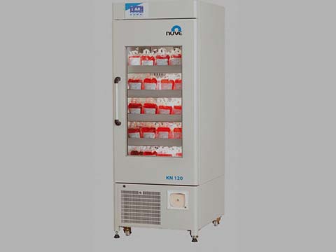
The securing of donated blood and other blood properties in the laboratory for the purpose of blood transfusion along with other medical practices is known as blood banking.

Examining the blood for any sign of a contagious sickness and tagging it for the blood transfer procedure is entailed.

In 2013, the American Association of Blood Banks postulated that:

  • Each day around 36,000 blood segments are in demand
  • Annually, around 13.6 million blood segments are donated
  • Per annual season, the blood donators are around 6.8 million in number

Some Blood cell parts,  types of which are erythrocytes, blood plasma, cryoprecipitated AHF, and thrombocytes are being disintegrated per blood segment.

Facts to know about Blood donation

Various sufferers with necessities which vary are being injected once with a detached full segment of blood. Additional 21 million parts of the blood are being injected.

Nevertheless, at times, in a situation where a sufferer is about to go through surgery  some weeks after, they might see the need to donate their blood for an emergency should the surgery require a blood transfusion.

The process in which you donate blood for yourself is known as an autologous donation. The benchmarks to be taken note of for willing blood donors are as follows:

  • Either in correspondence with the state law, or Ought to be aged 16,
  • Ought to be in a favorable state of health
  • Ought to be weighing at least 110 pounds on the scale
  • Before any donation is made, they have to perform well in the chronology health condition and bodily test
  • With the agreement on the guardians part, teenagers are allowed to be blood donors in some states.

In a blood banking, What are the experiments to be taken place?

Without limitations, immediately blood is donated the laboratory conducts a series of examination which are in no particular order.

  • Typing: ABO group (blood type)
  • Rh typing (positive or negative antigen)

In order not to cause harm to the blood recipient, the erythrocytes are being analyzed for any sign of disguised antibodies

The recipient body is tested for any sign of illness whether presently or formerly .

some examples of such illness may include:

  • Hepatitis viruses B and C
  • Human immunodeficiency virus (HIV)
  • Human T-lymphotropic viruses (HTLV) I and II
  • Syphilis
  • West Nile virus
  • Chagas disease

Irradiation to blood cells is carried out to disable any T-lymphocytes present in the donated blood. (T-lymphocytes can cause a response whilst transfused, however can also purpose graft-versus-host troubles with repeated publicity to foreign cells.)

Leukocyte-decreased blood has been filtered to remove the white blood cells that include antibodies which could cause fevers inside the recipient of the transfusion. (These antibodies, with repeated transfusions, might also boom a recipient’s chance of reactions to subsequent transfusions.)

What Is a Blood Bank Refrigerator?

A blood bank refrigerator stores biomedical components like blood merchant at managed temperatures. For protection, the machine includes tracking equipment to regularly take a look at and report temperatures.

It may additionally have security features like locking doors to protect the quality of the contents. Blood banks and facilities that work with blood product might also want to use specialized refrigeration gadgets to stay compliant with the law and keep excessive storage requirements for affected person safety.

Units come in a range of sizes for distinctive packages and may be custom built if essential. Donated blood needs to be well saved before its transfused to an affected person in want.

One situation with storing blood products is the slender range of acceptable garage temperatures vital.

Essential facts about blood bank refrigerators and blood storage

If blood gets too warm or too cold, it is able to be spoiled, or there could be issues almost it’s intercity that might make it inadvisable to transfuse.

The blood bank fridge very cautiously controls temperatures and uses tight vacuum seals and enthusiasts to preserve the environment inside a slender temperature variety.

In addition, it could encompass rapid healing systems to equalize the temperature when the door is opened and closed.

Donors are commonly screened before their plasma or blood donations are taken.

For safety, some blood financial institution fridge equipment sounds an alarm or sends a signal to a tracking room while the temperature exceeds the secure range.

An operator can check the refrigerator, affirm that there may be a temperature abnormality, and deal with the problem.

Logs can be saved in digital layout to generate a document showing that the facility’s refrigerators were maintained within the right variety.

Donated blood should not get too warm or too cold.

Another trouble may be a bacterial infection. A blood bank refrigerator can be made from bacteria-resistant additives, with production that makes it clean to clean.

Sliding shelves and trays can be rounded so that they obtained the puncture weight of blood product, and can encompass grips with antibacterial plastic coatings to preserve the interior easily.

The blood bank refrigerator doors can also lock to control the right of gaining access to, limiting the probabilities of infection and waste. Locks might also open with keys or number pads, depending on layout and the extent of gaining access to manage needed.

In a few cases, cooled blood may be warmed before it’s given to a patient.

In addition to refrigeration gadgets ranging in size from beneath-counter units to few-door refrigerators, it’s additionally feasible to install a walk-in cooler.

For blood banks that take care of large volumes of clots, this can be necessary to offer sufficient room for storage. Blood bank freezers also are available for plasma, which can be frozen for storage and thawed when it is needed.

The pleasant alternative can depend on the type and extent of blood products handled in a facility.

What are the blood sorts?

According to the American Association of Blood Banks, distribution of blood sorts  within the U.S. Consists of the subsequent:

  • O Rh-positive – 39%
  • A Rh-positive – 31%
  • B Rh-positive – 9%
  • O Rh-negative – 9%
  • A Rh-negative – 6%
  • AB Rh-positive – 3%
  • B Rh-negative – 2%
  • AB Rh-negative – 1%

What are the components of blood?

While blood, or one in every of its components, may be transferred, each element serves many functions, which includes the following:

Red blood cells: These cells bring oxygen to the tissues in the body and are generally used inside the treatment of anemia.

Platelets:  They assist the blood to clot and are used within the treatment of leukemia and other categories of most cancers.

White blood cells: These cells assist to combat infection, and useful resource inside the immune method.

Plasma:  The watery, liquid part of the blood wherein the pink blood cells, white blood cells, and platelets are suspended. Plasma is needed to hold the numerous elements of the blood through the bloodstream. Plasma serves many functions, along with the following:

  • Helps to maintain blood stress
  • Provides proteins for blood clotting
  • Balances the tiers of sodium and potassium

Cryoprecipitate AHF. The part of the plasma that contains clotting factors that help to manipulate bleeding.

Albumin, immune globulins, and clotting factor concentrates can also be separated and processed for transfusions.

Recommendation

Types of sphygmomanometer

Blood Chemistry Analyzer

Best medical equipment for Doctors

Top Physical therapy Equipment for Home

Binocular microscope parts and functions

Binocular microscope parts and functions

Nzubechi Uchegbu | February 8th, 2022


Let us take a look at the Binocular microscope parts and functions. One of the concerns of scientific research is that so much of what is going on is invisible to the bare eye.

A binocular microscope is also known as a compound microscope it is an optical microscope with two eyepieces to seriously ease viewing and cut down on eye strain.

Most microscopes bought these days are binocular microscopes though the interaction between the two lenses can range relying on the microscope type.

Parts of a Binocular Microscope

The three primary, structural additives of a compound microscope are the pinnacle, base, and arm.

While the optical parts of a Binocular microscope encompass:

The eyepiece

This is where your eyes will be. If the microscope is binocular, use each eyepieces. With binocular microscopes, you almost constantly can regulate the width of the eyepieces to make certain they healthy the spacing of your eyes.

The eyepiece incorporates the eyepiece lens, one of the  lenses doing the actual magnifying in a compound microscope.

Carrying arm

When moving a microscope, although it is just a few inches, usually pick it up by means of the wearing arm.

Do NOT drag the microscope: pick out it up. The microscope may have rubber feet that prevent it from sliding, so if you attempt to tug it, it’s going to shake and vibrate and viable harm components.

Never pick out up the microscope by way of any element aside from the wearing arm. The different parts are typically an awful lot extra fragile and susceptible to breaking in case you attempt.

The objective lenses

Most compound mild microscopes will incorporate 3 to 4 goal lenses that can be circled over the slide. Sometimes these lenses are just known as objectives.

When a particular objective has been fully circled into role, you may pay attention or feel a click on as that objective locks into place.

The objective lens is the second one of the two lenses doing the actual magnifying in a compound microscope, so if it is not snapped into right position, you won’t see the proper image.

Each goal lens can generally be unscrewed from its position in the rotating turret that houses it. Be cautious you’re rotating the turret, not unscrewing an objective. Do NOT unscrew the goals from the turret.

Each goal lens has a specific magnifying power, so the photograph for your slide might be magnified to lesser or extra extents, relying on which objective lens you have got selected.

The magnification of a goal lens will continually be an entire number. There may be other things written on the side of an goal, however the one that is a whole variety more than 1 may be the magnification. You can forget about the entirety else written there.

The stage

The degree is the platform that the slide could be clipped on to.

Stage clips

The slide could be held in location on the stage with level clips. Most of the time, these will clip towards the perimeters of the slide. They do now not sit down above or under the slide.

Furthermore, they are spring-loaded to maintain the slide edges and lock the slide in vicinity in order that the degree controls can move the placement of the slide smoothly.

If the slide isn’t always clipped in area, you will not be able to reposition the slide to locate microscopic features of interest.

Stage Controls

These can help you pass your slide at the same time as you’re viewing it, but handiest if the slide is nicely clipped in with the stage clips.

Always discover wherein these are to your microscope earlier than you begin viewing your slide.

They appear to by no means be within the same area in  exclusive microscopes and in case you simply blindly grope for them whilst viewing your slide, you may possibly do something unfortunate for your view or to the entire microscope.

There are continually  dials. One movements the slide left and right. The different moves the slide up and down.

Sometimes they’re on top of every different, occasionally they’re  separate dials, occasionally they’re above the stage,  every so often they may be under the level.  Spend a few seconds to discover them each time earlier than you sit down down at a microscope.

Coarse attention

This is constantly the bigger of the 2 awareness knobs. You need to normally simplest want to apply the coarse attention knob as soon as for each new slide. Use it with the lowest power goal to get the specimen about in focus.

After that, most effective use the great awareness knob, even after you exchange to a better-power goal.

Sometimes the coarse recognition recognise is with the best awareness knob. Sometimes it’s far break away the first-rate focus knob.

Fine focus

This is usually the smaller of the 2 cognizance knobs. This is the focus know you’ll use over and over once more in viewing slides.

Don’t trade the coarse consciousness after the usage of it for the first time, simplest change the great focus.

Condenser adjustment

Not all microscopes have a condenser adjustment knob. If there are most effective  knobs,  the ones two are the coarse focus and the satisfactory consciousness and also you most effective need to maintain the ones two separate. But if there’s a 3rd knob, it is the condenser adjustment knob.

As a standard rule, do NOT touch or alter this knob. It controls how far the light condenser is from the slide, which must be well adjusted before you operate the microscope.

If you move it, you will have it in the wrong role. If your scope has the knob, discover in which it is and keep away from it.

Diaphragm

This is immediately below the hollow within the level wherein mild passes through to the slide. It is controlled by a degree which opens and closes an iris to let greater or less light via the slide.

In some specimens there is not much comparison among the colors and sun shades of the distinct additives being magnified.

Changing how vivid the view is by adjusting the diaphragm can allow you to better see a number of the information you are attempting to exaggerate.

Functions of parts of the Binocular Microscope

The parts of a Binocular or compound microscope are divided into Structural components and Optical components

Structural additives features:

  1. Head/Body homes the optical parts within the upper part of the microscope
  2. Base of the microscope supports the microscope and houses the illuminator
  3. Arm connects to the base and supports the microscope head. It is also used to carry the microscope.
  4. When wearing a compound microscope always take care to boost it by way of both the arm and base, simultaneously.

Optical components functions:

Eyepiece Lenses and Objective Lenses:

Eyepiece or Ocular is what you glance through at the pinnacle of the microscope. Typically, well known eyepieces have a magnifying power of 10x. Optional eyepieces of various powers are available, normally from 5x-30x.

Eyepiece Tube holds the eyepieces in region above the goal lens. Binocular microscope heads commonly incorporate a diopter adjustment ring that allows for the viable inconsistencies of our eyesight in a single or each eyes. The monocular (unmarried eye usage) microscope does now not need a diopter.

Binocular microscopes also swivel (Interpupillary Adjustment) to allow for exclusive distances between the eyes of various people.

Objective Lenses are the number one optical lenses on a microscope. They variety from 4x-100x and typically, consist of, three, 4 or five on lens on maximum microscopes. Objectives may be forward or rear-dealing with.

Nosepiece houses the targets. The objectives are uncovered and are installed on a rotating turret so that special objectives can be without problems selected. Standard objectives include 4x, 10x, 40x and 100x despite the fact that special energy targets are to be had.

Coarse and Fine Focus knobs are used to recognition the microscope. Increasingly, they are coaxial knobs – that is to mention they’re constructed on the same axis with the great focus knob at the out of doors. Coaxial awareness knobs are greater handy since the viewer does now not must grope for a exceptional knob.

Stage is where the specimen to be considered is positioned. A mechanical level is used whilst running at higher magnifications wherein delicate actions of the specimen slide are required.

Stage Clips are used while there’s no mechanical stage. The viewer is needed to transport the slide manually to view one-of-a-kind sections of the specimen.

Aperture is the hole inside the degree through which the base (transmitted) mild reaches the stage.

Illuminator is the mild source for a microscope, generally located in the base of the microscope. Most mild microscopes use low voltage, halogen bulbs with non-stop variable lights manipulate placed inside the base.

Condenser is used to acquire and cognizance the light from the illuminator directly to the specimen. It is placed beneath the degree regularly along with an iris diaphragm.

Iris Diaphragm controls the quantity of mild achieving the specimen. It is located above the condenser and underneath the stage.

Most excessive nice microscopes include an Abbe condenser with an iris diaphragm. Combined, they control both the focal point and quantity of light implemented to the specimen.

Condenser Focus Knob moves the condenser up or down to manipulate the lighting fixtures attention at the specimen.

Conclusion

The binocular microscope is one of the most critical gear utilized in chemistry and biology. This instrument lets in a scientist or physician to magnify an object to study it in element. I am confident you now know about the Binocular microscope parts and functions.

Many styles of microscopes exist, allowing distinct ranges of magnification and producing extraordinary varieties of image. Some of the maximum advanced microscopes may even see atoms.

Recommendations

Types of sphygmomanometer

List of home-use medical equipment

What is a cath lab? staff, equipment list, and procedure

uses of analytical balance

Best Uses of Analytical Balance

Dr. Chuwkuebuka | February 3rd, 2022


Do you know about the Uses of Analytical Balance? today everything you ought to know about analytical balance is unveiled.

Analytical balances are precision measuring instruments that are used in quantitative chemical analysis to determine the mass of solids, liquids, powders, and granular substances.

Electronic balances now use the magnetic force restoration principle, with readability up to.0001 g. Analytical scales or balances are classified as very common or laboratory instruments. Analytical scales must be present in all laboratories because they are one of the most frequently used tools.

Analytical scales fall into the category of general laboratory equipment under these conditions. Other tools in the category of general laboratory equipment include laminar airflow, moisture analyzers, and micropipettes, in addition to analytical scales.

Uses of an Analytical Balance

Analytical scales are typically used by laboratory workers. As a result, analytical scales are also known as laboratory scales. The people who use the scales in this laboratory are referred to as laboratory assistants.

These scales are commonly used by analysts and researchers, in addition to laboratory assistants.

Analytical scales are used to weigh the composition of new substances from a set of substances. The ORS solution is one such example.

A bad-tasting ORS solution can be consumed if it is flavored with the appropriate composition of guava. Using analytical scales, researchers can determine the weight composition that is appropriate for the mixture in order to achieve the best results.

Types of Analytical Balance

  • Analog Analytical Balance
  •  Digital Analytical Balance

Analog Analytical Balance

This type of analytical balance still uses a manual operation process. The operation is carried out by sliding the slider.

It is necessary for you to move the slider in order to see the mass of a substance using analog analytical scales.

 Digital Analytical Balance

Unlike analog analytical scales, digital analytical scales are simple to use. To calculate the mass of a substance, simply place the substance to be measured in the provided container, also known as the balance pan.

Digital analytical scales are popular among laboratory assistants and researchers because they are simple to use and allow them to work more practically and efficiently.

Analytical Balance Components

  1. A balance plate (pan) is a container in which a sample of material to be measured in mass is placed. You can clean this area with a special cleaning brush or a clean microfiber cloth.
  2. Weights are used to calibrate analytical scales. The weights’ mass is precisely calculated so that the balance calibration remains accurate.
  3. Passing water through a tool used to determine the position of the balance plate. The balance plate is also repositioned using Waterpass.

4.The power button (on / off button) is used to turn on or off the balance. After pressing the power button to turn on the balance, it is usually left idle for about 10-15 minutes before employing it to produce a more precise measurement

  1. The ‘Re-zero’ or ‘Tare’ button is used to reset the balance to a neutral position (zero).

It is recommended that you do not use this button too frequently so that the balance can continue to produce accurate measurements.

  1. The ‘Mode’ button is used to specify the conversion system to be used when measuring.

You can change the conversion system by clicking the ‘Mode’ button.

Methods for maintaining an Analytical Balance

The first method of caring for analytic scales is to place them on a table or flat section.

Analytical scales should not be placed in bumpy areas or in areas that are frequently exposed to interaction of gadgets or different human beings due to the fact analytical scales are touchy devices.

Another way is not to drop items which can be too heavy and intense in analytical scales. Especially for those excessive gadgets, commonly, it’s miles nevertheless inside the limits of the maximum size of analytical scales, but it’s miles still risky for the conditions of analytical scales for later use.

The next way is to not always use analytical scales for unsafe chemicals directly on pinnacle of analytical scales. We advise which you use other bins first as a place to position those dangerous chemical substances.

For materials or items that need to be weighed to have severe temperatures, along with too cold or too hot, you must wait some time for the item to be in a situation of regular temperature or room temperature.

Another way to treat analytical scales is to avoid the usage of brushes which can be too easy for cleansing analytical scales. In addition, do now not frequently move or shift analytical scales to diverse locations. Analytic scales is probably damaged and the effects of the scales are now not correct.

Approaches of the usage of an Analytical Balance

First, to start the usage of analytical scales within the laboratory, you have to take a seat effectively in the front of the analytical scales.

Don’t forget about to position your analytical balance in the front of you at a distance that you can without problems attain.

Happens, you’ll now not have difficulties whilst the usage of it. Observe the analytical scales you may use because there might not be any accustomed to the use of new analytical scales.

After that, you could start turning on the analytical scales utilized by pressing the power button. But before that, you need to first take a look at the electricity you operate. Have you used the energy supply or now not.

More encouraged whilst using analytical scales, use a strength supply in order that the electric strength generated may be solid.

Analytical scales are on. Then what to do? You need to wait first in order that the location of the variety on the display is solid, an alias is at zero.

Usually, in a few instances, the analytical scales used do not display 0. To be able to be in a solid quantity, then you definitely ought to appearance back to the guide so that you can do the settings first.

The subsequent step is to weigh. Usually, there are analytic scales which have a protector round them. For that, you ought to first open the protective door earlier than inserting the fabric or object to be weighed at the disk in it.

As defined above, analytical scales need to be used with caution. Likewise whilst putting substances or specific in it.

Do it carefully with the aid of considering the maximum limit of the potential of the analytical balance. If you do now not take note of the maximum limit, then the analytical balance used can be damaged.

After the fabric or item that you want to weigh is already on the analytical scale, do not right now record the numbers that come out at the screen.

That variety isn’t yet strong. It’s an awesome concept to wait a few moments a good way to observe how many masses of cloth or items you are weighing.

When you have got finished weighing, do now not go away any material or object that has been weighed in the analytical stability. Immediately put off it in order that the analytical stability is empty.

The next step is to clean analytical scales. The tool that can be used is a small brush. Clean analytical scales calmly to hold them clean after use.

Finally, the protective cowl of the analytical scales tightly so that it will preserve best and cleanliness.

Top Industries make use of Analytical Balance

The particular measurements offered by an analytical scale makes it a critical piece of equipment for any zone that is based on quantitative chemical evaluation.

They are to be had in various shapes and sizes and can show readouts in diverse gadgets, making them suitable for a huge variety of enterprise programs.

These encompass pharmaceutical, food, plastics and chemical manufacturing, quality assurance labs and calibration laboratories, and so forth.

Conclusion

As defined above, analytical scales do need to be used with warning. If not used cautiously and according with the situations, the ensuing scales can honestly be messy and now not according with reality. As such, analytical scales have to be used in unique rooms with the least quantity of disturbance.

These scales additionally need to be monitored carefully and are often calibrated. Most analytical scales have internal calibration and automated calibration with external weights.

Recommendations

Physical therapy equipment for home

Top Korean medical equipment manufacturers

types of Sphygmomanometers

Best types of Sphygmomanometers

Dr. Chuwkuebuka | January 27th, 2022


The different types of Sphygmomanometers would be thoroughly looked at since it is among the essential devices used in medical practice.

The modern lifestyle has made various fitness dangers inclusive of high blood pressure, coronary heart disease, and different common cardiovascular situations an increasing number of not unusual.

Picking the right sphygmomanometer is a smart desire that we all do not have to forget every day, for an ideal living.

What is a sphygmomanometer?

A sphygmomanometer is an instrument used to measure the degree of blood strain which is likewise called a blood stress meter or blood strain gauge or blood pressure monitor.

The word sphygmomanometer is derived from the Greek phrase ‘sphygmos’ which means beating of the heart or the pulse and manometer imply the tool used for measuring the pressure or anxiety.

This instrument was invented by Samuel Siegfried Karl Ritter von Basch in the year 1881. But in the year 1896, Scipione Riva-Rocci added a simplified version of the sphygmomanometer.

Advancements within the sphygmomanometer, or blood stress gauges as they’re also often known as, had been pretty impressive over time.

Medical devices have come to be extra specialized, and numerous styles of sphygmomanometers have become available in the market.

TYPES OF SPHYGMOMANOMETERS

Following are the three foremost varieties of sphygmomanometer:

  1. Mercury Sphygmomanometer
  2. Aneroid Sphygmomanometer
  3. Automatic Digital Sphygmomanometer
  • Mercury Sphygmomanometer

The mercury sphygmomanometer is the most conventional form of blood strain equipment, and it could be considered the golden standard inside the health enterprise.

Mercury sphygmomanometers are made of manually inflatable cuffs which might be connected to measuring gadgets with mercury-infused tubes.

While operating the device, it’s really crucial to place the apparatus on a flat floor and in an upright position to get the correct readings. These devices are very sensitive and require unique care, and if by chance dropped can cause a rupture of mercury, rendering the tool vain and doubtlessly risky.

The largest benefit of the usage of mercury sphygmomanometers is that they’re quite smooth to apply, and if used well, can remaining an entire life.

The device can produce the most accurate outcomes without requiring a good deal readjustment. Due to the toxic nature of its contents, but, using mercury sphygmomanometers has been banned in some international locations, and different kinds of sphygmomanometers are being used as an alternative.

  • Aneroid Sphygmomanometer

Aneroid way “without fluid,” and because the call suggests, this form of BP apparatus doesn’t make use of any mercury and is consequently taken into consideration the safest alternative to mercury sphygmomanometers.

Its recording strategies are quite just like that of the mercury sphygmomanometer, besides the stethoscope’s attachment to the cuff.

In the android tool, the cuff is attached to a dial gauge with tubing. The gauge head includes mechanical components that convert the cuff stress into gauge-based studying. Additionally, there are numerous other styles of aneroid sphygmomanometers relying on how they’re used.

These may include :

  • pocket-aneroid sphygmomanometers
  • palm aneroid sphygmomanometers
  • clock-fashion aneroid sphygmomanometers

Ultimately, the benefit of the usage of aneroid sphygmomanometers is the affordability of the apparatus, as well as its compact and accessible nature.

However, similar to other devices used to check the degree of blood pressure, aneroid sphygmomanometers employ a delicate mechanism and consequently necessitates careful coping with.

Aneroid sphygmomanometers additionally require recalibration with the aid of specialists, to keep away from cases of defective studying.

  • Automatic Digital Sphygmomanometer

The automatic digital sphygmomanometer is the maximum technologically superior sphygmomanometer design so far.

These devices use a digital stress sensor to measure blood pressure, and the readings are offered on a virtual show.

Similar to mercury and aneroid sphygmomanometers, digital sphygmomanometers also come with inflatable cuffs, however there may be a distinction within the manner for measuring blood strain.

Unlike the other two kinds, computerized virtual sphygmomanometers evaluate and measure the fluctuations of arteries. These gadgets are perfect for home use as they may be the easiest to perform.

To avoid the hazard of inaccurate readings, periodic counter tests with conventional mercury sphygmomanometers are endorsed.

FAQs on the types of Sphygmomanometers

Below are the most suitable answers to the FAQs on the about the various types of sphygmomanometer;

Are digital sphygmomanometer results accurate? Yes, the results produced by the digital sphygmomanometer are accurate however, repeating the test process is recommended for assurance and validation of the result.

What are the different Parts of sphygmomanometer?

Bladder:

This is an inflatable bag this is used to compress the arm to occlude the artery. To make sure complete arterial compression, bladders have to have specific sizing parameters.

Cuff:

This is designed to keep the bladder around the arm all through the dimension. For correct dimension, the cuff have to be designed properly with respect to placement and the placement.

Manometer:

This is a tool used to degree the air pressure in mmHg. The manometer used in an aneroid sphygmomanometer includes an eye-like motion to measure the air stress carried out to the cuff. To increase the diaphragm, the gauge has a series of copper or beryllium and there are gears to transform the linear movement of the diaphragm to get the readings in mmHg.

Valve:

This used as a deflation valve to control the cuff. This plays a essential role in getting an correct measurement.

Bulb:

This is used to pump the air into the cuff.

Among the manual and digital sphygmomanometer, which one is mostly recommended in hospitals? This is based on individual difference and preference, however, priority is mostly to the manual sphygmomanometer.

Why is Sphygmomanometers important for household? Monitoring one’s blood pressure is a obligation not just of medical doctors and physicians, but of each person alike.

Having abnormal blood stress degrees, whether or not improved or dropped, are positive caution signs and symptoms of ill fitness, and checking for these abnormalities is an critical step in the direction of the prevention of high blood pressure, stroke, or heart assault.

Therefore, households as lots as clinics and hospitals must spend money on the right sphygmomanometers for common use.

when was sphygmomanometer invented? It was invented in 1881 by von Basch and the device was the first non-invasive BP measurements.

Which arm to measure blood pressure right or left? This is the choice of your healthcare provider to make. Both arms can give an accurate reading of the blood pressure when the proper guides are considered.

How to Use Sphygmomanometers

In using sphygmomanometers, processes vary in step with the type and form of sphygmomanometer used, but all types perform on a single precept.

A bulb will increase the stress via inflating the cuff placed round one’s arm, and a valve releases the stress. During this technique, a stethoscope is used to concentrate to arterial blood glide sounds.

As the heartbeats, blood going through the arteries causes a upward push in strain, which speedy decreases while the heart’s ventricles prepare for some other beat. This upward thrust and fall are known as systolic and diastolic stress, respectively.

Steps for accurate blood pressure measurement

Below are the essential steps to  follow in order appropriately degree one’s blood stress:

  • Know your simple expertise of blood pressure readings

Blood strain readings are expressed as systolic over diastolic stress. The regular blood strain of a median healthy grownup is 120/80.

  • Use proper-sized cuffs

Cuffs which can be too loose or too tight might also influence the accuracy of blood pressure measurements. The cuff have to be eighty% of the circumference of the upper arm. Be sure not to tighten the cuff on a clothed arm.

Properly tight the cuff at the arm. While wrapping the cuff around the higher arm, preserve the lower edge of the cuff one inch above the antecubital fossa, the area of the arm before the elbow.

Position the stethoscope. Lightly press the stethoscope bell over the brachial artery, simply under the decrease fringe of the cuff.

Inflate the cuff. Using the bulb, inflate the cuff to a hundred and eighty mmHg, then launch the usage of the valve at a mild rate of about 3 mm consistent with 2nd.

  • Listen and watch carefully

While examining the dial or mercury gauge, concentrate for a knocking sound with the stethoscope.

The first knocking sound represents the systolic blood strain of the patient. The point when the sound disappears in the meantime indicates the diastolic stress.

  • Repeat the manner on the opposite arm

For correct evaluation, it’s far encouraged that the blood pressure of both hands be measured. The difference in size of both palms, in addition to the patient’s position and cuff length, should be mentioned.

How to properly choose the right sphyg for you

Below is a list of the applicable elements one must recall earlier when selecting what sphygmomanometer to purchase.

  • Needs and Price

The right sphygmomanometer is determined by the wishes and necessities of its consumer. Aneroid sphygmomanometers provide readings of excessive accuracy but require enough know-how and information of coping with the tool.

Nurses, physicians, and experts should, consequently, search for durable, bendy, and excessive-accuracy devices consisting of these, which could resist heavy use.

Individuals searching to shop for sphygmomanometers for private use, in the meantime, may also take into account shopping sphygmomanometers which can be simplest to perform a number of the many types.

Digital sphygmomanometers are designed to be very consumer-friendly, especially for people who do no longer have any medical background. Price is any other determining component for what sphygmomanometer to purchase, and the allocated price range can fantastically slender down one’s decision.

The price variety of a respectable sphygmomanometer varies from 10 to 70 bucks, relying on the sort. Devices that fall within the better fee tiers are those designed for specialists who require excessive-acting units.

  • Accuracy

As with any clinical device, the accuracy of measurements is of extreme importance.

As mentioned in advance, the accuracy of sphygmomanometers in large part depends on its type–mercury sphygmomanometers provide the same old of measurements and is therefore of maximum accuracy; aneroid sphygmomanometers also offer readings of very excessive accuracy, however require sufficient knowledge and expertise of handling the device; digital sphygmomanometers generally tend to sacrifice convenience and simplicity of use for accuracy of measurements, compared to the opposite  sphygmomanometer types.

  • Quality, Design, and Cuff Size

Sphygmomanometers vary in line with their exceptional of cloth and design, all of which have an effect on the overall overall performance of the tool. Therefore, all components of a sphygmomanometer need to be of pinnacle-notch quality.

The cuff fabric, gauge, inflation bulb, and valve should all ideally be well constructed, non-sticking, and hypoallergenic.

A perfect gauge must have three hundred mmHg of pressure, and the bulb element should be constructed from latex-loose fabric.

For a sphygmomanometer, cuff length is also extraordinarily critical. Cuff sizes that might be both too unfastened- or tight-fit would possibly bring about wrong readings.

Therefore, one needs to make sure that the width of the sphygmomanometer cuff presents a size variety that could match its user perfectly, whether for private or scientific use.

For reference, 80% of the arm needs to be blanketed by using the air bladder of the cuff. Small cuff sizes are for arm diameters of 17 to 22 cm, medium cuff sizes are for 22 to 32 cm, and big cuff sizes are for 33 to 42 cm arm diameters. For medical experts, it’s far cautioned to buy a medium-sized cuff that can be adjusted to healthy sufferers’ larger and smaller sizes.

  • Convenience and Portability

Sphygmomanometers, whether or not used regularly and in one-of-a-kind places or bought for domestic use, has to be evaluated primarily based on convenience and simplicity of use.

Those who are looking to buy a sphygmomanometer specifically for domestic use can choose to keep a couple of dollars and select a tool that isn’t lightweight or pocket-sized.

Final thought

The importance of this device for hospitals, organizations, and households can never be over-emphasized. Your heart state and other health complications can be easily detected when the blood pressure is known.

Most times, heart diseases like hypertension is known as a silent killer and can only be detected using different types of sphygmomanometers. Always consult your healthcare provider while choosing the right sphygmomanometer suitable for you.

Recommendations

List of top use medical devices

Best medical equipment for doctors